


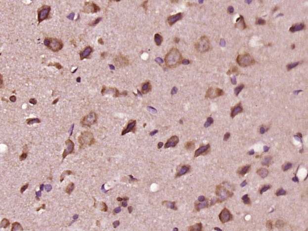
Immunohistochemical staining of human stomach shows moderate cytoplasmic positivity in glandular cells.
Immunohistochemical staining of human stomach shows moderate cytoplasmic positivity in glandular cells.
Anti-EDEM3 Antibody
HPA025755
ApplicationsImmunoHistoChemistry
Product group Antibodies
ReactivityHuman
TargetEDEM3
Overview
- SupplierAtlas Antibodies
- Product NameAnti-EDEM3 Antibody
- Delivery Days Customer4
- ApplicationsImmunoHistoChemistry
- CertificationResearch Use Only
- ClonalityPolyclonal
- ConjugateUnconjugated
- Gene ID80267
- Target nameEDEM3
- Target descriptionER degradation enhancing alpha-mannosidase like protein 3
- Target synonymsC1orf22, CDG2V, ER degradation-enhancing alpha-mannosidase-like protein 3, ER degradation enhancer, mannosidase alpha-like 3, ER degradation-enhancing -mannosidase-like protein 3, ER degradation-enhancing alpha-mannosidase-like 3, alpha-1,2-mannosidase EDEM3
- HostRabbit
- IsotypeIgG
- Protein IDQ9BZQ6
- Protein NameER degradation-enhancing alpha-mannosidase-like protein 3
- Scientific DescriptionRecombinant Protein Epitope Signature Tag (PrEST) antigen sequence
- ReactivityHuman
- Storage Instruction-20°C,2°C to 8°C
- UNSPSC41116161
Datasheet
MSDS
Related products
Product group Antibodies
EDEM3 Polyclonal AntibodyBS-14502R
ApplicationsImmunoFluorescence, ELISA, ImmunoCytoChemistry, ImmunoHistoChemistry, ImmunoHistoChemistry Frozen, ImmunoHistoChemistry Paraffin
ReactivityBovine, Chicken, Equine, Human, Mouse, Porcine, Rat, Sheep
TargetEDEM3
- SizePrice
Product group Antibodies
Anti-EDEM3 (C-term) Antibody102-22351
ApplicationsWestern Blot
TargetEDEM3
- SizePrice
Product group Antibodies

EDEM3 AntibodyLS-C780037
ApplicationsWestern Blot, ELISA
ReactivityHuman, Mouse
TargetEDEM3
- SizePrice

Product group Antibodies
Anti-EDEM3 AntibodyHPA025754
ApplicationsImmunoHistoChemistry
ReactivityHuman
TargetEDEM3
- SizePrice

Product group Antibodies
Anti-EDEM3 AntibodyHPA027297
ApplicationsImmunoCytoChemistry
ReactivityHuman
TargetEDEM3
- SizePrice

Product group Antibodies
Anti-EDEM3 Antibody Picoband(r)A10297-2-CARRIER-FREE
ApplicationsWestern Blot, ELISA, ImmunoHistoChemistry
ReactivityHuman
TargetEDEM3
- SizePrice