


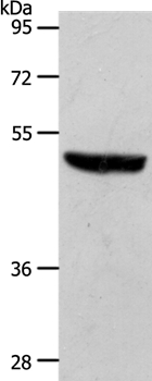
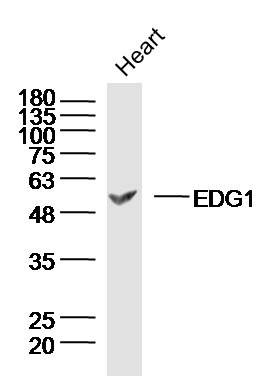
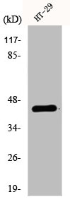
Figure 1. Western blot analysis of EDG1 using anti-EDG1 antibody (M01502). Electrophoresis was performed on a 5-20% SDS-PAGE gel at 70V (Stacking gel) / 90V (Resolving gel) for 2-3 hours. The sample well of each lane was loaded with 30 ug of sample under reducing conditions. Lane 1: human HepG2 whole cell lysates, Lane 2: human U-87MG whole cell lysates, Lane 3: human SH-SY5Y whole cell lysates, Lane 4: human U251 whole cell lysates, Lane 5: rat brain tissue lysates, Lane 6: rat C6 whole cell lysates, Lane 7: mouse brain tissue lysates, Lane 8: mouse Neuro-2a whole cell lysates. After electrophoresis, proteins were transferred to a nitrocellulose membrane at 150 mA for 50-90 minutes. Blocked the membrane with 5% non-fat milk/TBS for 1.5 hour at RT. The membrane was incubated with rabbit anti-EDG1 antigen affinity purified monoclonal antibody (Catalog # M01502) at 1:500 overnight at 4°C, then washed with TBS-0.1%Tween 3 times with 5 minutes each and probed with a goat anti-rabbit IgG-HRP secondary antibody at a dilution of 1:500 for 1.5 hour at RT. The signal is developed using an Enhanced Chemiluminescent detection (ECL) kit (Catalog # EK1002) with Tanon 5200 system. A specific band was detected for EDG1 at approximately 43 kDa. The expected band size for EDG1 is at 43 kDa.
Figure 1. Western blot analysis of EDG1 using anti-EDG1 antibody (M01502). Electrophoresis was performed on a 5-20% SDS-PAGE gel at 70V (Stacking gel) / 90V (Resolving gel) for 2-3 hours. The sample well of each lane was loaded with 30 ug of sample under reducing conditions. Lane 1: human HepG2 whole cell lysates, Lane 2: human U-87MG whole cell lysates, Lane 3: human SH-SY5Y whole cell lysates, Lane 4: human U251 whole cell lysates, Lane 5: rat brain tissue lysates, Lane 6: rat C6 whole cell lysates, Lane 7: mouse brain tissue lysates, Lane 8: mouse Neuro-2a whole cell lysates. After electrophoresis, proteins were transferred to a nitrocellulose membrane at 150 mA for 50-90 minutes. Blocked the membrane with 5% non-fat milk/TBS for 1.5 hour at RT. The membrane was incubated with rabbit anti-EDG1 antigen affinity purified monoclonal antibody (Catalog # M01502) at 1:500 overnight at 4°C, then washed with TBS-0.1%Tween 3 times with 5 minutes each and probed with a goat anti-rabbit IgG-HRP secondary antibody at a dilution of 1:500 for 1.5 hour at RT. The signal is developed using an Enhanced Chemiluminescent detection (ECL) kit (Catalog # EK1002) with Tanon 5200 system. A specific band was detected for EDG1 at approximately 43 kDa. The expected band size for EDG1 is at 43 kDa.
Anti-EDG1 S1PR1 Rabbit Monoclonal Antibody
M01502
ApplicationsWestern Blot
Product group Antibodies
ReactivityHuman, Mouse, Rat
TargetS1PR1
Overview
- SupplierBoster Bio
- Product NameAnti-EDG1 S1PR1 Rabbit Monoclonal Antibody
- Delivery Days Customer9
- ApplicationsWestern Blot
- CertificationResearch Use Only
- ClonalityMonoclonal
- Clone IDAABO-19
- Gene ID1901
- Target nameS1PR1
- Target descriptionsphingosine-1-phosphate receptor 1
- Target synonymsCD363, CHEDG1, D1S3362, ECGF1, EDG-1, EDG1, S1P1, sphingosine 1-phosphate receptor 1, S1P receptor Edg-1, endothelial differentiation G-protein coupled receptor 1, endothelial differentiation, sphingolipid G-protein-coupled receptor, 1, sphingosine 1-phosphate receptor EDG1, sphingosine 1-phosphate receptor Edg-1
- HostRabbit
- IsotypeIgG
- Protein IDP21453
- Protein NameSphingosine 1-phosphate receptor 1
- Scientific DescriptionBoster Bio Anti-EDG1 S1PR1 Rabbit Monoclonal Antibody catalog # M01502. Tested in WB application. This antibody reacts with Human, Mouse, Rat.
- ReactivityHuman, Mouse, Rat
- Storage Instruction-20°C
- UNSPSC12352203
References
- Cao R, Sun R, Ye Y, et al. Low shear stress-induced blockage of autophagic flux impairs endothelial barrier and facilitates atherosclerosis in mice. Exp Cell Res. 2024,439(1):114071. doi: 10.1016/j.yexcr.2024.114071Read this paper
Datasheet
MSDS
Related products
Product group Antibodies
Anti-S1PR1 Antibody144-12935
ApplicationsWestern Blot
ReactivityHuman, Mouse
TargetS1PR1
- SizePrice
Product group Antibodies
Anti-S1PR1 AntibodyA43229
ApplicationsWestern Blot
ReactivityHuman, Mouse
- SizePrice
Product group Antibodies
References
EDG1 Polyclonal AntibodyBS-7112R
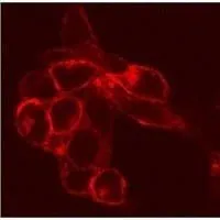
ApplicationsImmunoFluorescence, Western Blot, ELISA, ImmunoCytoChemistry, ImmunoHistoChemistry, ImmunoHistoChemistry Frozen, ImmunoHistoChemistry Paraffin
ReactivityCanine, Equine, Human, Mouse, Rat
TargetS1PR1
- SizePrice
Product group Antibodies
S1PR1 AntibodyCSB-PA002247
ApplicationsImmunoFluorescence, Western Blot, ELISA
ReactivityHuman, Mouse, Rat
TargetS1PR1
- SizePrice
Product group Antibodies
S1PR1 Polyclonal AntibodyCAC13921
ApplicationsImmunoFluorescence, Western Blot, ELISA, ImmunoHistoChemistry
ReactivityMouse
TargetS1PR1
- SizePrice
Product group Antibodies

S1PR1 / EDG1 / S1P1 AntibodyLS-C402837
ApplicationsWestern Blot, ELISA
ReactivityHuman, Mouse, Rat
TargetS1PR1
- SizePrice

Product group Antibodies
Anti-S1PR1 AntibodyHPA075568
ApplicationsImmunoCytoChemistry
ReactivityHuman
TargetS1PR1
- SizePrice
Product group Antibodies
EDG1 antibodyGTX11424
ApplicationsFlow Cytometry, ImmunoFluorescence, ImmunoPrecipitation, Western Blot, ImmunoCytoChemistry, ImmunoHistoChemistry, ImmunoHistoChemistry Frozen
ReactivityHuman, Mouse, Rat
TargetS1PR1
- SizePrice
Product group Antibodies
Anti-EDG1/HEXIM1 AntibodyCAB12935
ApplicationsWestern Blot, ELISA
ReactivityHuman
TargetS1PR1
- SizePrice