


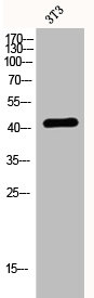
Figure 1. Western blot analysis of EDG3 using anti-EDG3 antibody (M03755-1). Electrophoresis was performed on a 5-20% SDS-PAGE gel at 70V (Stacking gel) / 90V (Resolving gel) for 2-3 hours. The sample well of each lane was loaded with 30 ug of sample under reducing conditions. Lane 1: human HepG2 whole cell lysates, Lane 2: human Hela whole cell lysates. After electrophoresis, proteins were transferred to a nitrocellulose membrane at 150 mA for 50-90 minutes. Blocked the membrane with 5% non-fat milk/TBS for 1.5 hour at RT. The membrane was incubated with rabbit anti-EDG3 antigen affinity purified monoclonal antibody (Catalog # M03755-1) at 1:1000 overnight at 4°C, then washed with TBS-0.1%Tween 3 times with 5 minutes each and probed with a goat anti-rabbit IgG-HRP secondary antibody at a dilution of 1:500 for 1.5 hour at RT. The signal is developed using an Enhanced Chemiluminescent detection (ECL) kit (Catalog # EK1002) with Tanon 5200 system. A specific band was detected for EDG3 at approximately 42 kDa. The expected band size for EDG3 is at 42 kDa.
Figure 1. Western blot analysis of EDG3 using anti-EDG3 antibody (M03755-1). Electrophoresis was performed on a 5-20% SDS-PAGE gel at 70V (Stacking gel) / 90V (Resolving gel) for 2-3 hours. The sample well of each lane was loaded with 30 ug of sample under reducing conditions. Lane 1: human HepG2 whole cell lysates, Lane 2: human Hela whole cell lysates. After electrophoresis, proteins were transferred to a nitrocellulose membrane at 150 mA for 50-90 minutes. Blocked the membrane with 5% non-fat milk/TBS for 1.5 hour at RT. The membrane was incubated with rabbit anti-EDG3 antigen affinity purified monoclonal antibody (Catalog # M03755-1) at 1:1000 overnight at 4°C, then washed with TBS-0.1%Tween 3 times with 5 minutes each and probed with a goat anti-rabbit IgG-HRP secondary antibody at a dilution of 1:500 for 1.5 hour at RT. The signal is developed using an Enhanced Chemiluminescent detection (ECL) kit (Catalog # EK1002) with Tanon 5200 system. A specific band was detected for EDG3 at approximately 42 kDa. The expected band size for EDG3 is at 42 kDa.
Anti-EDG3 Rabbit Monoclonal Antibody
M03755-1
ApplicationsImmunoFluorescence, Western Blot, ImmunoCytoChemistry, ImmunoHistoChemistry
Product group Antibodies
ReactivityHuman
TargetS1PR3
Overview
- SupplierBoster Bio
- Product NameAnti-EDG3 Rabbit Monoclonal Antibody
- Delivery Days Customer9
- ApplicationsImmunoFluorescence, Western Blot, ImmunoCytoChemistry, ImmunoHistoChemistry
- CertificationResearch Use Only
- ClonalityMonoclonal
- Clone ID25S21
- Gene ID1903
- Target nameS1PR3
- Target descriptionsphingosine-1-phosphate receptor 3
- Target synonymsC9orf108, C9orf47, EDG-3, EDG3, LPB3, S1P3, bA791O21.3, sphingosine 1-phosphate receptor 3, G protein-coupled receptor, endothelial differentiation gene-3, S1P receptor 3, S1P receptor EDG3, S1P receptor Edg-3, endothelial differentiation G-protein coupled receptor 3, endothelial differentiation, sphingolipid G-protein-coupled receptor, 3, sphingosine 1-phosphate receptor Edg-3, uncharacterized protein C9orf47
- HostRabbit
- IsotypeIgG
- Protein IDQ99500
- Protein NameSphingosine 1-phosphate receptor 3
- Scientific DescriptionBoster Bio Anti-EDG3 Rabbit Monoclonal Antibody catalog # M03755-1. Tested in WB, IHC, ICC/IF applications. This antibody reacts with Human.
- ReactivityHuman
- Storage Instruction-20°C
- UNSPSC12352203
References
- Zhao T, Ding T, Sun Z, et al. SPHK1/S1P/S1PR pathway promotes the progression of peritoneal fibrosis by mesothelial-mesenchymal transition. FASEB J. 2024,38(2):e23417. doi: 10.1096/fj.202301323RRead this paper
Related products
Product group Antibodies
S1PR3 AntibodyCSB-PA002248
ApplicationsImmunoFluorescence, Western Blot, ELISA
ReactivityHuman
TargetS1PR3
- SizePrice

Product group Antibodies
Anti-EDG3 AntibodyA100005
ApplicationsImmunoFluorescence, Western Blot, ELISA
ReactivityHuman
- SizePrice
Product group Antibodies

Goat anti-EDG3EB09356
ApplicationsELISA, ImmunoHistoChemistry
ReactivityHuman
TargetS1PR3
- SizePrice
Product group Antibodies

SIPR3 / EDG3 / S1P3 AntibodyLS-C402838
ApplicationsWestern Blot, ELISA
ReactivityHuman, Mouse
TargetS1PR3
- SizePrice
Product group Antibodies
References
EDG3 Polyclonal AntibodyBS-7541R
ApplicationsFlow Cytometry, ImmunoFluorescence, Western Blot, ELISA, ImmunoCytoChemistry, ImmunoHistoChemistry, ImmunoHistoChemistry Frozen, ImmunoHistoChemistry Paraffin
ReactivityBovine, Human, Mouse, Rabbit, Rat
TargetS1PR3
- SizePrice
Product group Antibodies
SIPR3 antibodyGTX77925
ApplicationsImmunoHistoChemistry, ImmunoHistoChemistry Paraffin
ReactivityHuman
TargetS1PR3
- SizePrice
Product group Antibodies
Anti-EDG3 (N-term) Antibody102-26956
ApplicationsWestern Blot
TargetS1PR3
- SizePrice