


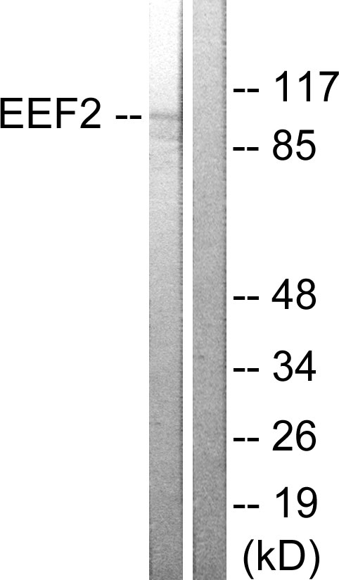
Figure 1. Western blot analysis of EEF2 using anti-EEF2 antibody (A00830-1). Electrophoresis was performed on a 5-20% SDS-PAGE gel at 70V (Stacking gel) / 90V (Resolving gel) for 2-3 hours. The sample well of each lane was loaded with 30 ug of sample under reducing conditions. Lane 1: human Jurkat whole cell lysates, Lane 2: monkey COS-7 whole cell lysates, Lane 3: human Hela whole cell lysates, Lane 4: human 293T whole cell lysates, Lane 5: human HepG2 whole cell lysates, Lane 6: human Daudi whole cell lysates, Lane 7: human MCF-7 whole cell lysates, Lane 8: zebrafish tissue lysates, Lane 9: rat stomach tissue lysates, Lane 10: rat pancrease tissue lysates, Lane 11: rat C6 whole cell lysates, Lane 12: mouse pancrease tissue lysates, Lane 13: mouse 3T3L1 whole cell lysates. After electrophoresis, proteins were transferred to a nitrocellulose membrane at 150 mA for 50-90 minutes. Blocked the membrane with 5% non-fat milk/TBS for 1.5 hour at RT. The membrane was incubated with rabbit anti-EEF2 antigen affinity purified polyclonal antibody (Catalog # A00830-1) at 0.25 microg/mL overnight at 4°C, then washed with TBS-0.1%Tween 3 times with 5 minutes each and probed with a goat anti-rabbit IgG-HRP secondary antibody at a dilution of 1:5000 for 1.5 hour at RT. The signal is developed using an Enhanced Chemiluminescent detection (ECL) kit (Catalog # EK1002) with Tanon 5200 system. A specific band was detected for EEF2 at approximately 95 kDa. The expected band size for EEF2 is at 95 kDa.
. EEF2 was detected in a paraffin-embedded section of human breast cancer tissue. Heat mediated antigen retrieval was performed in EDTA buffer (pH 8.0, epitope retrieval solution). The tissue section was blocked with 10% goat serum. The tissue section was then incubated with 2 microg/ml rabbit anti-EEF2 Antibody (A00830-1) overnight at 4°C. Peroxidase Conjugated Goat Anti-rabbit IgG was used as secondary antibody and incubated for 30 minutes at 37°C. The tissue section was developed using HRP Conjugated Rabbit IgG Super Vision Assay Kit (Catalog # SV0002) with DAB as the chromogen.")
. EEF2 was detected in a paraffin-embedded section of human laryngeal squamous cell carcinoma tissue. Heat mediated antigen retrieval was performed in EDTA buffer (pH 8.0, epitope retrieval solution). The tissue section was blocked with 10% goat serum. The tissue section was then incubated with 2 microg/ml rabbit anti-EEF2 Antibody (A00830-1) overnight at 4°C. Peroxidase Conjugated Goat Anti-rabbit IgG was used as secondary antibody and incubated for 30 minutes at 37°C. The tissue section was developed using HRP Conjugated Rabbit IgG Super Vision Assay Kit (Catalog # SV0002) with DAB as the chromogen.")
. EEF2 was detected in a paraffin-embedded section of human lymphoma tissue. Heat mediated antigen retrieval was performed in EDTA buffer (pH 8.0, epitope retrieval solution). The tissue section was blocked with 10% goat serum. The tissue section was then incubated with 2 microg/ml rabbit anti-EEF2 Antibody (A00830-1) overnight at 4°C. Peroxidase Conjugated Goat Anti-rabbit IgG was used as secondary antibody and incubated for 30 minutes at 37°C. The tissue section was developed using HRP Conjugated Rabbit IgG Super Vision Assay Kit (Catalog # SV0002) with DAB as the chromogen.")
. EEF2 was detected in a paraffin-embedded section of human renal cancer tissue. Heat mediated antigen retrieval was performed in EDTA buffer (pH 8.0, epitope retrieval solution). The tissue section was blocked with 10% goat serum. The tissue section was then incubated with 2 microg/ml rabbit anti-EEF2 Antibody (A00830-1) overnight at 4°C. Peroxidase Conjugated Goat Anti-rabbit IgG was used as secondary antibody and incubated for 30 minutes at 37°C. The tissue section was developed using HRP Conjugated Rabbit IgG Super Vision Assay Kit (Catalog # SV0002) with DAB as the chromogen.")
. EEF2 was detected in a paraffin-embedded section of mouse ovary tissue. Heat mediated antigen retrieval was performed in EDTA buffer (pH 8.0, epitope retrieval solution). The tissue section was blocked with 10% goat serum. The tissue section was then incubated with 2 microg/ml rabbit anti-EEF2 Antibody (A00830-1) overnight at 4°C. Peroxidase Conjugated Goat Anti-rabbit IgG was used as secondary antibody and incubated for 30 minutes at 37°C. The tissue section was developed using HRP Conjugated Rabbit IgG Super Vision Assay Kit (Catalog # SV0002) with DAB as the chromogen.")
. EEF2 was detected in a paraffin-embedded section of rat ovary tissue. Heat mediated antigen retrieval was performed in EDTA buffer (pH 8.0, epitope retrieval solution). The tissue section was blocked with 10% goat serum. The tissue section was then incubated with 2 microg/ml rabbit anti-EEF2 Antibody (A00830-1) overnight at 4°C. Peroxidase Conjugated Goat Anti-rabbit IgG was used as secondary antibody and incubated for 30 minutes at 37°C. The tissue section was developed using HRP Conjugated Rabbit IgG Super Vision Assay Kit (Catalog # SV0002) with DAB as the chromogen.")
Figure 1. Western blot analysis of EEF2 using anti-EEF2 antibody (A00830-1). Electrophoresis was performed on a 5-20% SDS-PAGE gel at 70V (Stacking gel) / 90V (Resolving gel) for 2-3 hours. The sample well of each lane was loaded with 30 ug of sample under reducing conditions. Lane 1: human Jurkat whole cell lysates, Lane 2: monkey COS-7 whole cell lysates, Lane 3: human Hela whole cell lysates, Lane 4: human 293T whole cell lysates, Lane 5: human HepG2 whole cell lysates, Lane 6: human Daudi whole cell lysates, Lane 7: human MCF-7 whole cell lysates, Lane 8: zebrafish tissue lysates, Lane 9: rat stomach tissue lysates, Lane 10: rat pancrease tissue lysates, Lane 11: rat C6 whole cell lysates, Lane 12: mouse pancrease tissue lysates, Lane 13: mouse 3T3L1 whole cell lysates. After electrophoresis, proteins were transferred to a nitrocellulose membrane at 150 mA for 50-90 minutes. Blocked the membrane with 5% non-fat milk/TBS for 1.5 hour at RT. The membrane was incubated with rabbit anti-EEF2 antigen affinity purified polyclonal antibody (Catalog # A00830-1) at 0.25 microg/mL overnight at 4°C, then washed with TBS-0.1%Tween 3 times with 5 minutes each and probed with a goat anti-rabbit IgG-HRP secondary antibody at a dilution of 1:5000 for 1.5 hour at RT. The signal is developed using an Enhanced Chemiluminescent detection (ECL) kit (Catalog # EK1002) with Tanon 5200 system. A specific band was detected for EEF2 at approximately 95 kDa. The expected band size for EEF2 is at 95 kDa.
Anti-EEF2 Antibody Picoband(r)
A00830-1-CARRIER-FREE
ApplicationsWestern Blot, ELISA, ImmunoHistoChemistry
Product group Antibodies
ReactivityHuman, Monkey, Mouse, Rat, Zebra Fish
TargetEEF2
Overview
- SupplierBoster Bio
- Product NameAnti-EEF2 Antibody Picoband(r)
- Delivery Days Customer9
- ApplicationsWestern Blot, ELISA, ImmunoHistoChemistry
- CertificationResearch Use Only
- ClonalityPolyclonal
- Concentration500 ug/ml
- Gene ID1938
- Target nameEEF2
- Target descriptioneukaryotic translation elongation factor 2
- Target synonymsEEF-2, EF-2, EF2, SCA26, elongation factor 2, epididymis secretory sperm binding protein, polypeptidyl-tRNA translocase
- HostRabbit
- IsotypeIgG
- Protein IDP13639
- Protein NameElongation factor 2
- Scientific DescriptionBoster Bio Anti-EEF2 Antibody Picoband® catalog # A00830-1. Tested in ELISA, IHC, WB applications. This antibody reacts with Human, Monkey, Mouse, Rat, Zebrafish. The brand Picoband indicates this is a premium antibody that guarantees superior quality, high affinity, and strong signals with minimal background in Western blot applications. Only our best-performing antibodies are designated as Picoband, ensuring unmatched performance.
- ReactivityHuman, Monkey, Mouse, Rat, Zebra Fish
- Storage Instruction-20°C,2°C to 8°C
- UNSPSC12352203
Related products
Product group Antibodies
Anti-eEF2 AntibodyA95374
ApplicationsImmunoFluorescence, Western Blot, ELISA, ImmunoHistoChemistry
ReactivityHuman, Mouse, Rat
- SizePrice
Product group Antibodies
Anti-EEF2 Antibody144-02068
ApplicationsWestern Blot
ReactivityHuman
TargetEEF2
- SizePrice
Product group Antibodies
EEF2 Recombinant Antibody, AbBy Fluor-350 ConjugatedBSM-61650R-BF350
ApplicationsFlow Cytometry, ImmunoFluorescence, Western Blot
ReactivityHuman, Mouse, Rat
TargetEEF2
- SizePrice
Product group Antibodies
EEF2 AntibodyCSB-PA002260
ApplicationsImmunoFluorescence, Western Blot, ELISA, ImmunoHistoChemistry
ReactivityHuman, Mouse, Rat
TargetEEF2
- SizePrice
Product group Antibodies
Eef2 Polyclonal AntibodyCAC07503
ApplicationsImmunoFluorescence, ELISA, ImmunoHistoChemistry
TargetEEF2
- SizePrice
Product group Antibodies
eEF2 antibody [C1C3]GTX102286
ApplicationsWestern Blot
ReactivityHuman
TargetEEF2
- SizePrice

Product group Antibodies
Anti-EEF2 AntibodyHPA040534
ApplicationsWestern Blot, ImmunoCytoChemistry, ImmunoHistoChemistry
ReactivityHuman
TargetEEF2
- SizePrice
Product group Antibodies

EEF2 / Elongation Factor 2 AntibodyLS-C763468
ApplicationsImmunoHistoChemistry
ReactivityHuman
TargetEEF2
- SizePrice