


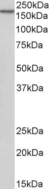
Figure 1. Western blot analysis of EGFR using anti-EGFR antibody (A00023-4). Electrophoresis was performed on a 5-20% SDS-PAGE gel at 70V (Stacking gel) / 90V (Resolving gel) for 2-3 hours. The sample well of each lane was loaded with 50ug of sample under reducing conditions. Lane 1: human placenta tissue lysates, Lane 2: human U87 whole cell lysates, Lane 3: human A431 whole cell lysates, Lane 4: human A549 whole cell lysates, Lane 5: human Hela whole cell lysates, Lane 6: rat liver tissue lysates, Lane 7: mouse liver tissue lysates. After Electrophoresis, proteins were transferred to a Nitrocellulose membrane at 150mA for 50-90 minutes. Blocked the membrane with 5% Non-fat Milk/ TBS for 1.5 hour at RT. The membrane was incubated with rabbit anti-EGFR antigen affinity purified polyclonal antibody (Catalog # A00023-4) at 0.25 microg/mL overnight at 4°C, then washed with TBS-0.1%Tween 3 times with 5 minutes each and probed with a goat anti-rabbit IgG-HRP secondary antibody at a dilution of 1:10000 for 1.5 hour at RT. The signal is developed using an Enhanced Chemiluminescent detection (ECL) kit (Catalog # EK1002) with Tanon 5200 system. A specific band was detected for EGFR at approximately 180KD. The expected band size for EGFR is at 180KD.
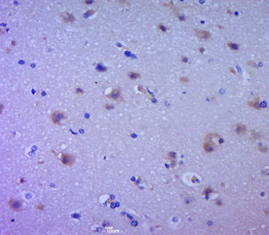
. EGFR was detected in paraffin-embedded section of human lung cancer tissue. Heat mediated antigen retrieval was performed in EDTA buffer (pH8.0, epitope retrieval solution). The tissue section was blocked with 10% goat serum. The tissue section was then incubated with 1microg/ml rabbit anti-EGFR Antibody (A00023-4) overnight at 4°C. Biotinylated goat anti-rabbit IgG was used as secondary antibody and incubated for 30 minutes at 37°C. The tissue section was developed using Strepavidin-Biotin-Complex (SABC) (Catalog # SA1022) with DAB as the chromogen.")
. EGFR was detected in paraffin-embedded section of human mammary cancer tissue. Heat mediated antigen retrieval was performed in EDTA buffer (pH8.0, epitope retrieval solution). The tissue section was blocked with 10% goat serum. The tissue section was then incubated with 1microg/ml rabbit anti-EGFR Antibody (A00023-4) overnight at 4°C. Biotinylated goat anti-rabbit IgG was used as secondary antibody and incubated for 30 minutes at 37°C. The tissue section was developed using Strepavidin-Biotin-Complex (SABC) (Catalog # SA1022) with DAB as the chromogen.")
. EGFR was detected in paraffin-embedded section of mouse liver tissue. Heat mediated antigen retrieval was performed in EDTA buffer (pH8.0, epitope retrieval solution). The tissue section was blocked with 10% goat serum. The tissue section was then incubated with 1microg/ml rabbit anti-EGFR Antibody (A00023-4) overnight at 4°C. Biotinylated goat anti-rabbit IgG was used as secondary antibody and incubated for 30 minutes at 37°C. The tissue section was developed using Strepavidin-Biotin-Complex (SABC) (Catalog # SA1022) with DAB as the chromogen.")
. EGFR was detected in paraffin-embedded section of rat liver tissue. Heat mediated antigen retrieval was performed in EDTA buffer (pH8.0, epitope retrieval solution). The tissue section was blocked with 10% goat serum. The tissue section was then incubated with 1microg/ml rabbit anti-EGFR Antibody (A00023-4) overnight at 4°C. Biotinylated goat anti-rabbit IgG was used as secondary antibody and incubated for 30 minutes at 37°C. The tissue section was developed using Strepavidin-Biotin-Complex (SABC) (Catalog # SA1022) with DAB as the chromogen.")
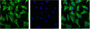
. EGFR was detected in paraffin-embedded section of human lung cancer tissue. Heat mediated antigen retrieval was performed in EDTA buffer (pH8.0, epitope retrieval solution). The tissue section was blocked with 10% goat serum. The tissue section was then incubated with 4microg/mL rabbit anti-EGFR Antibody (A00023-4) overnight at 4°C. DyLight®488 Conjugated Goat Anti-Rabbit IgG (BA1127) was used as secondary antibody at 1:100 dilution and incubated for 30 minutes at 37°C. The section was counterstained with DAPI. Visualize using a fluorescence microscope and filter sets appropriate for the label used.")
. EGFR was detected in immunocytochemical section of A431 cells. Enzyme antigen retrieval was performed using IHC enzyme antigen retrieval reagent (AR0022) for 15 mins. The cells were blocked with 10% goat serum. And then incubated with 4microg/mL rabbit anti-EGFR Antibody (A00023-4) overnight at 4°C. DyLight®550 Conjugated Goat Anti-Rabbit IgG (BA1135) was used as secondary antibody at 1:100 dilution and incubated for 30 minutes at 37°C. The section was counterstained with DAPI. Visualize using a fluorescence microscope and filter sets appropriate for the label used.")
. Overlay histogram showing A431 cells stained with A00023-4 (Blue line).To facilitate intracellular staining, cells were fixed with 4% paraformaldehyde and permeabilized with permeabilization buffer. The cells were blocked with 10% normal goat serum. And then incubated with rabbit anti-EGFR Antibody (A00023-4,1microg/1x106 cells) for 30 min at 20°C. DyLight®488 conjugated goat anti-rabbit IgG (BA1127, 5-10microg/1x106 cells) was used as secondary antibody for 30 minutes at 20°C. Isotype control antibody (Green line) was rabbit IgG (1microg/1x106) used under the same conditions. Unlabelled sample without incubation with primary antibody and secondary antibody (Red line) was used as a blank control.")
Figure 1. Western blot analysis of EGFR using anti-EGFR antibody (A00023-4). Electrophoresis was performed on a 5-20% SDS-PAGE gel at 70V (Stacking gel) / 90V (Resolving gel) for 2-3 hours. The sample well of each lane was loaded with 50ug of sample under reducing conditions. Lane 1: human placenta tissue lysates, Lane 2: human U87 whole cell lysates, Lane 3: human A431 whole cell lysates, Lane 4: human A549 whole cell lysates, Lane 5: human Hela whole cell lysates, Lane 6: rat liver tissue lysates, Lane 7: mouse liver tissue lysates. After Electrophoresis, proteins were transferred to a Nitrocellulose membrane at 150mA for 50-90 minutes. Blocked the membrane with 5% Non-fat Milk/ TBS for 1.5 hour at RT. The membrane was incubated with rabbit anti-EGFR antigen affinity purified polyclonal antibody (Catalog # A00023-4) at 0.25 microg/mL overnight at 4°C, then washed with TBS-0.1%Tween 3 times with 5 minutes each and probed with a goat anti-rabbit IgG-HRP secondary antibody at a dilution of 1:10000 for 1.5 hour at RT. The signal is developed using an Enhanced Chemiluminescent detection (ECL) kit (Catalog # EK1002) with Tanon 5200 system. A specific band was detected for EGFR at approximately 180KD. The expected band size for EGFR is at 180KD.
Anti-EGFR Antibody Picoband(r)
A00023-4-CARRIER-FREE
ApplicationsFlow Cytometry, ImmunoFluorescence, Western Blot, ELISA, ImmunoCytoChemistry, ImmunoHistoChemistry
Product group Antibodies
ReactivityHuman, Mouse, Rat
TargetEGFR
Overview
- SupplierBoster Bio
- Product NameAnti-EGFR Antibody Picoband(r)
- Delivery Days Customer9
- Application Supplier NoteTested Species: In-house tested species with positive results. By Heat: Boiling the paraffin sections in 10mM citrate buffer, pH6.0, for 20mins is required for the staining of formalin/paraffin sections. Other applications have not been tested. Optimal dilutions should be determined by end users.
- ApplicationsFlow Cytometry, ImmunoFluorescence, Western Blot, ELISA, ImmunoCytoChemistry, ImmunoHistoChemistry
- CertificationResearch Use Only
- ClonalityPolyclonal
- Concentration500 ug/ml
- Gene ID1956
- Target nameEGFR
- Target descriptionepidermal growth factor receptor
- Target synonymsERBB, ERBB1, ERRP, HER1, NISBD2, NNCIS, PIG61, mENA, epidermal growth factor receptor, EGFR vIII, avian erythroblastic leukemia viral (v-erb-b) oncogene homolog, cell growth inhibiting protein 40, cell proliferation-inducing protein 61, epidermal growth factor receptor tyrosine kinase domain, erb-b2 receptor tyrosine kinase 1, proto-oncogene c-ErbB-1, receptor tyrosine-protein kinase erbB-1
- HostRabbit
- IsotypeIgG
- Protein IDP00533
- Protein NameEpidermal growth factor receptor
- Scientific DescriptionBoster Bio Anti-EGFR Antibody Picoband® catalog # A00023-4. Tested in ELISA, Flow Cytometry, IF, IHC, ICC, WB applications. This antibody reacts with Human, Mouse, Rat. The brand Picoband indicates this is a premium antibody that guarantees superior quality, high affinity, and strong signals with minimal background in Western blot applications. Only our best-performing antibodies are designated as Picoband, ensuring unmatched performance.
- ReactivityHuman, Mouse, Rat
- Storage Instruction-20°C,2°C to 8°C
- UNSPSC12352203
Related products
Product group Antibodies
Anti-EGFR AntibodyA83204
ApplicationsWestern Blot, ELISA
ReactivityHuman
- SizePrice
Product group Antibodies

Anti-EGFR [C225 (Cetuximab)]Ab00279-1.1
ApplicationsFlow Cytometry, Neutralisation/Blocking
ReactivityHuman
TargetEGFR
- SizePrice
Product group Antibodies
ApplicationsFlow Cytometry
TargetEGFR
- SizePrice
Product group Antibodies
Anti-EGFR (N-term) Antibody131-0167
ApplicationsELISA
ReactivityHuman
TargetEGFR
- SizePrice
Product group Antibodies

EGFR-S1026 AntibodyABX032521
ApplicationsFlow Cytometry, Western Blot, ELISA, ImmunoHistoChemistry
- SizePrice

Product group Antibodies
Anti-EGFR AntibodyAMAB90816
ApplicationsWestern Blot, ImmunoHistoChemistry
ReactivityHuman
TargetEGFR
- SizePrice
Product group Antibodies
EGFR Polyclonal AntibodyBS-0165R
ApplicationsFlow Cytometry, ImmunoFluorescence, Western Blot, ELISA, ImmunoCytoChemistry, ImmunoHistoChemistry, ImmunoHistoChemistry Frozen, ImmunoHistoChemistry Paraffin
ReactivityCanine, Human, Mouse, Porcine, Rat
TargetEGFR
- SizePrice
Product group Antibodies
EGFR Monoclonal AntibodyCSB-MA000199
ApplicationsImmunoFluorescence, Western Blot, ELISA, ImmunoHistoChemistry
ReactivityHuman
TargetEGFR
- SizePrice
Product group Antibodies
References

Goat anti-EGFREB06962
ApplicationsWestern Blot, ELISA
ReactivityHuman
TargetEGFR
- SizePrice