


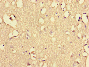
Immunohistochemical staining of human kidney shows strong membranous positivity in cells in glomeruli.
Immunohistochemical staining of human kidney shows strong membranous positivity in cells in glomeruli.
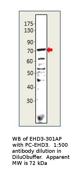
Anti-EHD3 Antibody
HPA049986
ApplicationsImmunoHistoChemistry
Product group Antibodies
ReactivityHuman
TargetEHD3
Overview
- SupplierAtlas Antibodies
- Product NameAnti-EHD3 Antibody
- Delivery Days Customer4
- ApplicationsImmunoHistoChemistry
- CertificationResearch Use Only
- ClonalityPolyclonal
- ConjugateUnconjugated
- Gene ID30845
- Target nameEHD3
- Target descriptionEH domain containing 3
- Target synonymsPAST3, EH domain-containing protein 3, PAST homolog 3
- HostRabbit
- IsotypeIgG
- Protein IDQ9NZN3
- Protein NameEH domain-containing protein 3
- Scientific DescriptionRecombinant Protein Epitope Signature Tag (PrEST) antigen sequence
- ReactivityHuman
- Storage Instruction-20°C,2°C to 8°C
- UNSPSC41116161
Datasheet
MSDS
Related products
Product group Antibodies
Anti-EHD3 AntibodyA54995
ApplicationsWestern Blot, ELISA
ReactivityHuman, Mouse, Rat
- SizePrice
Product group Antibodies
EHD3 AntibodyPACO47114
ApplicationsImmunoFluorescence, ELISA, ImmunoHistoChemistry
ReactivityHuman
TargetEHD3
- SizePrice
Product group Antibodies

EHD3 Antibody (Biotin)LS-C682179
ApplicationsELISA
ReactivityHuman
TargetEHD3
- SizePrice

Product group Antibodies
Anti-EHD3 AntibodyHPA049890
ApplicationsWestern Blot, ImmunoHistoChemistry
ReactivityHuman
TargetEHD3
- SizePrice
Product group Antibodies
EHD3 AntibodyCSB-PA878940LA01HU
ApplicationsImmunoFluorescence, ELISA, ImmunoHistoChemistry
ReactivityHuman
TargetEHD3
- SizePrice

Product group Antibodies
Anti-EHD3 Antibody Picoband(r)A04576-1-CARRIER-FREE
ApplicationsFlow Cytometry, ImmunoFluorescence, Western Blot, ImmunoCytoChemistry, ImmunoHistoChemistry
ReactivityHuman, Monkey, Mouse, Rat
TargetEHD3
- SizePrice