


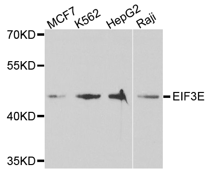
Figure 1. Western blot analysis of EIF3E using anti-EIF3E antibody (M00481-1). Electrophoresis was performed on a 5-20% SDS-PAGE gel at 70V (Stacking gel) / 90V (Resolving gel) for 2-3 hours. The sample well of each lane was loaded with 30 ug of sample under reducing conditions. Lane 1: human Raji whole cell lysates, Lane 2: human K562 whole cell lysates, Lane 3: rat thymus tissue lysates, Lane 4: mouse thymus tissue lysates, Lane 5: mouse ANA-1 whole cell lysates, Lane 6: mouse SP2/0 whole cell lysates. After electrophoresis, proteins were transferred to a nitrocellulose membrane at 150 mA for 50-90 minutes. Blocked the membrane with 5% non-fat milk/TBS for 1.5 hour at RT. The membrane was incubated with mouse anti-EIF3E antigen affinity purified monoclonal antibody (Catalog # M00481-1) at 0.5 microg/mL overnight at 4°C, then washed with TBS-0.1%Tween 3 times with 5 minutes each and probed with a goat anti-mouse IgG-HRP secondary antibody at a dilution of 1:10000 for 1.5 hour at RT. The signal is developed using an Enhanced Chemiluminescent detection (ECL) kit (Catalog # EK1001) with Tanon 5200 system. A specific band was detected for EIF3E at approximately 52 kDa. The expected band size for EIF3E is at 52 kDa.
. Overlay histogram showing U20S cells stained with M00481-1 (Blue line). To facilitate intracellular staining, cells were fixed with 4% paraformaldehyde and permeabilized with permeabilization buffer. The cells were blocked with 10% normal goat serum. And then incubated with mouse anti-EIF3E Antibody (M00481-1, 1 microg/1x106 cells) for 30 min at 20°C. DyLight®488 conjugated goat anti-mouse IgG (BA1126, 5-10 microg/1x106 cells) was used as secondary antibody for 30 minutes at 20°C. Isotype control antibody (Green line) was mouse IgG (1 microg/1x106) used under the same conditions. Unlabelled sample without incubation with primary antibody and secondary antibody (Red line) was used as a blank control.")
Figure 1. Western blot analysis of EIF3E using anti-EIF3E antibody (M00481-1). Electrophoresis was performed on a 5-20% SDS-PAGE gel at 70V (Stacking gel) / 90V (Resolving gel) for 2-3 hours. The sample well of each lane was loaded with 30 ug of sample under reducing conditions. Lane 1: human Raji whole cell lysates, Lane 2: human K562 whole cell lysates, Lane 3: rat thymus tissue lysates, Lane 4: mouse thymus tissue lysates, Lane 5: mouse ANA-1 whole cell lysates, Lane 6: mouse SP2/0 whole cell lysates. After electrophoresis, proteins were transferred to a nitrocellulose membrane at 150 mA for 50-90 minutes. Blocked the membrane with 5% non-fat milk/TBS for 1.5 hour at RT. The membrane was incubated with mouse anti-EIF3E antigen affinity purified monoclonal antibody (Catalog # M00481-1) at 0.5 microg/mL overnight at 4°C, then washed with TBS-0.1%Tween 3 times with 5 minutes each and probed with a goat anti-mouse IgG-HRP secondary antibody at a dilution of 1:10000 for 1.5 hour at RT. The signal is developed using an Enhanced Chemiluminescent detection (ECL) kit (Catalog # EK1001) with Tanon 5200 system. A specific band was detected for EIF3E at approximately 52 kDa. The expected band size for EIF3E is at 52 kDa.
Anti-EIF3e Antibody Picoband(r) (monoclonal, 10F5H6)
M00481-1-CY3
ApplicationsFlow Cytometry, Western Blot
Product group Antibodies
ReactivityHuman, Mouse, Rat
TargetEIF3E
Overview
- SupplierBoster Bio
- Product NameAnti-EIF3e Antibody Picoband(r) (monoclonal, 10F5H6)
- Delivery Days Customer9
- ApplicationsFlow Cytometry, Western Blot
- CertificationResearch Use Only
- ClonalityMonoclonal
- Clone ID10F5H6
- Concentration500 ug/ml
- ConjugateCy3
- Gene ID3646
- Target nameEIF3E
- Target descriptioneukaryotic translation initiation factor 3 subunit E
- Target synonymsEIF3-P48, EIF3S6, INT6, eIF3-p46, eukaryotic translation initiation factor 3 subunit E, eIF-3 p48, eukaryotic translation initiation factor 3 subunit 6, eukaryotic translation initiation factor 3, subunit 6 (48kD), eukaryotic translation initiation factor 3, subunit 6 48kDa, mammary tumor-associated protein INT6, murine mammary tumor integration site 6 (oncogene homolog), viral integration site protein INT-6 homolog
- HostMouse
- IsotypeIgG2b
- Protein IDP60228
- Protein NameEukaryotic translation initiation factor 3 subunit E
- Scientific DescriptionBoster Bio Anti-EIF3e Antibody Picoband® (monoclonal, 10F5H6) catalog # M00481-1. Tested in Flow Cytometry, WB applications. This antibody reacts with Human, Mouse, Rat. The brand Picoband indicates this is a premium antibody that guarantees superior quality, high affinity, and strong signals with minimal background in Western blot applications. Only our best-performing antibodies are designated as Picoband, ensuring unmatched performance.
- ReactivityHuman, Mouse, Rat
- Storage Instruction-20°C,2°C to 8°C
- UNSPSC12352203
Related products
Product group Antibodies
eIF3E Recombinant AntibodyBSM-62327R
ApplicationsImmunoFluorescence, Western Blot, ImmunoCytoChemistry
ReactivityHuman, Mouse, Rat
TargetEIF3E
- SizePrice
Product group Antibodies
Anti-EIF3E AntibodyA30972
ApplicationsImmunoFluorescence, Western Blot, ImmunoHistoChemistry
ReactivityHuman, Mouse, Rat
- SizePrice
Product group Antibodies
Anti-EIF3E Antibody144-05447
ApplicationsImmunoFluorescence, Western Blot
ReactivityHuman, Mouse
TargetEIF3E
- SizePrice
Product group Antibodies

Goat anti-EIF3E AntibodyEB10524
ApplicationsWestern Blot, ELISA
ReactivityBovine, Canine, Human, Porcine
TargetEIF3E
- SizePrice
Product group Antibodies
eIF3e antibodyGTX112342
ApplicationsImmunoFluorescence, Western Blot, ImmunoCytoChemistry, ImmunoHistoChemistry, ImmunoHistoChemistry Paraffin
ReactivityHuman, Mouse
TargetEIF3E
- SizePrice
Product group Antibodies

EIF3E AntibodyLS-C346085
ApplicationsImmunoFluorescence, Western Blot, ImmunoHistoChemistry
ReactivityHuman, Mouse
TargetEIF3E
- SizePrice

Product group Antibodies
Anti-EIF3E-25ulHPA023973
ApplicationsWestern Blot, ImmunoCytoChemistry, ImmunoHistoChemistry
ReactivityHuman, Mouse, Rat
- SizePrice
Product group Antibodies
EIF3E AntibodyCSB-PA007534ESR1HU
ApplicationsELISA, ImmunoHistoChemistry
ReactivityHuman
TargetEIF3E
- SizePrice