


Immunofluorescent staining of human cell line SiHa shows localization to nucleoplasm.
Immunofluorescent staining of human cell line SiHa shows localization to nucleoplasm.
Anti-ELK1 Antibody
HPA064381
ApplicationsImmunoCytoChemistry
Product group Antibodies
ReactivityHuman
TargetELK1
Overview
- SupplierAtlas Antibodies
- Product NameAnti-ELK1 Antibody
- Delivery Days Customer4
- ApplicationsImmunoCytoChemistry
- CertificationResearch Use Only
- ClonalityPolyclonal
- ConjugateUnconjugated
- Gene ID2002
- Target nameELK1
- Target descriptionETS transcription factor ELK1
- Target synonymsETS domain-containing protein Elk-1, ELK1, ETS transcription factor, ELK1, member of ETS oncogene family, ETS-like gene 1, tyrosine kinase (ELK1) oncogene
- HostRabbit
- IsotypeIgG
- Protein IDP19419
- Protein NameETS domain-containing protein Elk-1
- Scientific DescriptionRecombinant Protein Epitope Signature Tag (PrEST) antigen sequence
- ReactivityHuman
- Storage Instruction-20°C,2°C to 8°C
- UNSPSC41116161
Datasheet
MSDS
Related products
Product group Antibodies
Anti-ELK1 Antibody144-61648
ApplicationsWestern Blot, ImmunoHistoChemistry
ReactivityHuman, Mouse, Rat
TargetELK1
- SizePrice
Product group Antibodies
ApplicationsWestern Blot, ImmunoHistoChemistry
ReactivityHuman, Mouse, Rat
- SizePrice

Product group Antibodies
Anti-ELK1 Antibody Picoband(r)A01426-1-CARRIER-FREE
ApplicationsFlow Cytometry, Western Blot, ELISA
ReactivityHuman, Mouse, Rat
TargetELK1
- SizePrice
Product group Antibodies
ELK1 Polyclonal AntibodyCAC14532
ApplicationsWestern Blot, ELISA, ImmunoHistoChemistry
ReactivityMouse
TargetELK1
- SizePrice
Product group Antibodies
ELK1 AntibodyCSB-PA002319
ApplicationsImmunoFluorescence, Western Blot, ELISA, ImmunoHistoChemistry
ReactivityHuman, Mouse, Rat
TargetELK1
- SizePrice
Product group Antibodies
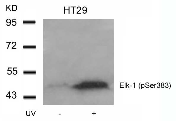
ELK1 (phospho Ser383) antibodyGTX11848
ApplicationsWestern Blot
ReactivityHuman, Mouse, Rat
TargetELK1
- SizePrice

Product group Antibodies
Anti-ELK1 AntibodyHPA036084
ApplicationsImmunoCytoChemistry, ImmunoHistoChemistry
ReactivityHuman
TargetELK1
- SizePrice
Product group Antibodies
Anti-ELK1 AntibodyHPA036084
ApplicationsImmunoCytoChemistry, ImmunoHistoChemistry
ReactivityHuman
TargetELK1
- SizePrice