


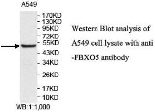
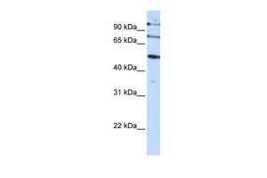
Figure 1. Western blot analysis of Emi1 using anti-Emi1 antibody (M05229-2). Electrophoresis was performed on a 5-20% SDS-PAGE gel at 70V (Stacking gel) / 90V (Resolving gel) for 2-3 hours. The sample well of each lane was loaded with 30 ug of sample under reducing conditions. Lane 1: human HepG2 whole cell lysates, Lane 2: human 293T whole cell lysates, Lane 3: human Hela whole cell lysates, Lane 4: human K562 whole cell lysates. After electrophoresis, proteins were transferred to a nitrocellulose membrane at 150 mA for 50-90 minutes. Blocked the membrane with 5% non-fat milk/TBS for 1.5 hour at RT. The membrane was incubated with rabbit anti-Emi1 antigen affinity purified monoclonal antibody (Catalog # M05229-2) at 1:500 overnight at 4°C, then washed with TBS-0.1%Tween 3 times with 5 minutes each and probed with a goat anti-rabbit IgG-HRP secondary antibody at a dilution of 1:500 for 1.5 hour at RT. The signal is developed using an Enhanced Chemiluminescent detection (ECL) kit (Catalog # EK1002) with Tanon 5200 system. A specific band was detected for Emi1 at approximately 65 kDa. The expected band size for Emi1 is at 50 kDa.
Figure 1. Western blot analysis of Emi1 using anti-Emi1 antibody (M05229-2). Electrophoresis was performed on a 5-20% SDS-PAGE gel at 70V (Stacking gel) / 90V (Resolving gel) for 2-3 hours. The sample well of each lane was loaded with 30 ug of sample under reducing conditions. Lane 1: human HepG2 whole cell lysates, Lane 2: human 293T whole cell lysates, Lane 3: human Hela whole cell lysates, Lane 4: human K562 whole cell lysates. After electrophoresis, proteins were transferred to a nitrocellulose membrane at 150 mA for 50-90 minutes. Blocked the membrane with 5% non-fat milk/TBS for 1.5 hour at RT. The membrane was incubated with rabbit anti-Emi1 antigen affinity purified monoclonal antibody (Catalog # M05229-2) at 1:500 overnight at 4°C, then washed with TBS-0.1%Tween 3 times with 5 minutes each and probed with a goat anti-rabbit IgG-HRP secondary antibody at a dilution of 1:500 for 1.5 hour at RT. The signal is developed using an Enhanced Chemiluminescent detection (ECL) kit (Catalog # EK1002) with Tanon 5200 system. A specific band was detected for Emi1 at approximately 65 kDa. The expected band size for Emi1 is at 50 kDa.
Anti-Emi1 Rabbit Monoclonal Antibody
M05229-2
ApplicationsImmunoFluorescence, ImmunoPrecipitation, Western Blot, ImmunoCytoChemistry, ImmunoHistoChemistry
Product group Antibodies
ReactivityHuman, Mouse, Rat
TargetFBXO5
Overview
- SupplierBoster Bio
- Product NameAnti-Emi1 Rabbit Monoclonal Antibody
- Delivery Days Customer9
- ApplicationsImmunoFluorescence, ImmunoPrecipitation, Western Blot, ImmunoCytoChemistry, ImmunoHistoChemistry
- CertificationResearch Use Only
- ClonalityMonoclonal
- Clone ID19F06
- Gene ID26271
- Target nameFBXO5
- Target descriptionF-box protein 5
- Target synonymsEMI1, FBX5, Fbxo31, F-box only protein 5, F-box protein Fbx5, early fission inhibitory protein 1, early mitotic inhibitor 1
- HostRabbit
- IsotypeIgG
- Protein IDQ9UKT4
- Protein NameF-box only protein 5
- Scientific DescriptionBoster Bio Anti-Emi1 Rabbit Monoclonal Antibody catalog # M05229-2. Tested in WB, IHC, ICC/IF, IP applications. This antibody reacts with Human, Mouse, Rat.
- ReactivityHuman, Mouse, Rat
- Storage Instruction-20°C
- UNSPSC12352203
Related products
Product group Antibodies
Anti-FBXO5 AntibodyA49628
ApplicationsWestern Blot, ELISA
ReactivityHuman, Mouse, Rat
- SizePrice
Product group Antibodies
Anti-EMI1 Antibody188-10380
ApplicationsFlow Cytometry
ReactivityHuman
TargetFBXO5
- SizePrice
Product group Antibodies
FBXO5 Recombinant AntibodyBSM-61974R
ApplicationsImmunoFluorescence, ImmunoPrecipitation, Western Blot, ImmunoCytoChemistry, ImmunoHistoChemistry, ImmunoHistoChemistry Frozen, ImmunoHistoChemistry Paraffin
ReactivityHuman, Mouse, Rat
TargetFBXO5
- SizePrice
Product group Antibodies
FBXO5 AntibodyCSB-PA008514GA01HU
ApplicationsWestern Blot, ELISA, ImmunoHistoChemistry
ReactivityHuman
TargetFBXO5
- SizePrice

Product group Antibodies
Anti-FBXO5 AntibodyHPA029048
ApplicationsWestern Blot, ImmunoCytoChemistry, ImmunoHistoChemistry
ReactivityHuman
TargetFBXO5
- SizePrice
Product group Antibodies
EMI1 antibody, C-termGTX46926
ApplicationsWestern Blot
ReactivityHuman
TargetFBXO5
- SizePrice
Product group Antibodies

FBXO5 / EMI1 AntibodyLS-C665549
ApplicationsWestern Blot, ELISA, ImmunoHistoChemistry
ReactivityHuman
TargetFBXO5
- SizePrice