


Immunofluorescent staining of human cell line U-2 OS shows localization to mitochondria.
Immunofluorescent staining of human cell line U-2 OS shows localization to mitochondria.
Anti-EXOG Antibody
HPA071050
ApplicationsImmunoCytoChemistry
Product group Antibodies
ReactivityHuman
TargetEXOG
Overview
- SupplierAtlas Antibodies
- Product NameAnti-EXOG Antibody
- Delivery Days Customer4
- ApplicationsImmunoCytoChemistry
- CertificationResearch Use Only
- ClonalityPolyclonal
- ConjugateUnconjugated
- Gene ID9941
- Target nameEXOG
- Target descriptionexo/endonuclease G
- Target synonymsENDOGL1, ENDOGL2, ENGL, ENGL-a, ENGL-b, ENGLA, ENGLB, nuclease EXOG, mitochondrial, endo G-like 1, endo/exonuclease (5'-3'), endonuclease G-like, endonuclease G-like 1, endonuclease G-like 2
- HostRabbit
- IsotypeIgG
- Protein IDQ9Y2C4
- Protein NameNuclease EXOG, mitochondrial
- Scientific DescriptionRecombinant Protein Epitope Signature Tag (PrEST) antigen sequence
- ReactivityHuman
- Storage Instruction-20°C,2°C to 8°C
- UNSPSC41116161
Datasheet
MSDS
Related products
Product group Antibodies
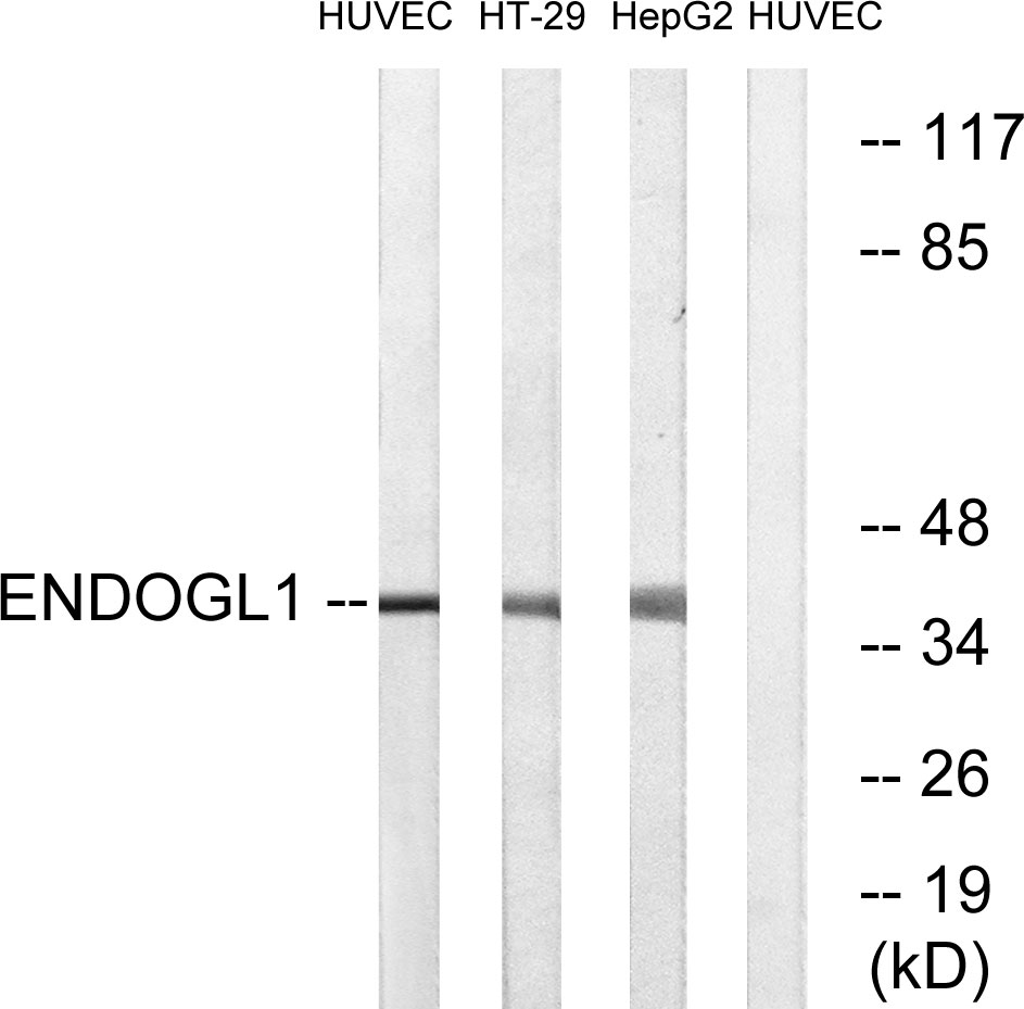
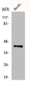
Anti-ENDOGL1 AntibodyA100167
ApplicationsWestern Blot, ELISA
ReactivityHuman
- SizePrice

Product group Antibodies
ApplicationsImmunoPrecipitation, Western Blot
ReactivityHuman
TargetEXOG
- SizePrice
Product group Antibodies
ApplicationsFlow Cytometry, Western Blot, ImmunoCytoChemistry
ReactivityHuman
TargetEXOG
- SizePrice
Product group Antibodies
EXOG AntibodyCSB-PA002341
ApplicationsWestern Blot, ELISA
ReactivityHuman
TargetEXOG
- SizePrice
Product group Antibodies

Goat anti-EXOGEB12764
ApplicationsWestern Blot, ELISA, ImmunoHistoChemistry
ReactivityHuman
TargetEXOG
- SizePrice
Product group Antibodies

EXOG / ENDOGL1 AntibodyLS-C335332
ApplicationsWestern Blot
ReactivityHuman
TargetEXOG
- SizePrice