

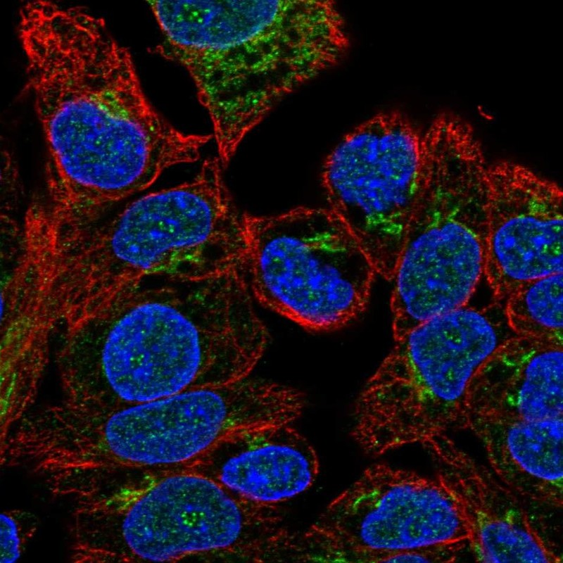
Immunofluorescent staining of human cell line Hep G2 shows localization to nucleus & the Golgi apparatus.
Immunofluorescent staining of human cell line Hep G2 shows localization to nucleus & the Golgi apparatus.
Anti-F5 Antibody
HPA002036
ApplicationsImmunoCytoChemistry
Product group Antibodies
ReactivityHuman
TargetF5
Overview
- SupplierAtlas Antibodies
- Product NameAnti-F5 Antibody
- Delivery Days Customer4
- ApplicationsImmunoCytoChemistry
- CertificationResearch Use Only
- ClonalityPolyclonal
- ConjugateUnconjugated
- Gene ID2153
- Target nameF5
- Target descriptioncoagulation factor V
- Target synonymsFVL, PCCF, RPRGL1, THPH2, coagulation factor V, activated protein c cofactor, coagulation factor V (proaccelerin, labile factor), coagulation factor V jinjiang A2 domain, factor V Leiden
- HostRabbit
- IsotypeIgG
- Protein IDP12259
- Protein NameCoagulation factor V
- Scientific DescriptionRecombinant Protein Epitope Signature Tag (PrEST) antigen sequence
- ReactivityHuman
- Storage Instruction-20°C,2°C to 8°C
- UNSPSC41116161
Datasheet
MSDS
Related products
Product group Antibodies

F5 / Factor Va Antibody (FITC)LS-C677871
ApplicationsELISA
ReactivityHuman
TargetF5
- SizePrice

Product group Antibodies
Anti-Factor V/F5 Antibody Picoband(r)A00440-1-CARRIER-FREE
ApplicationsWestern Blot
ReactivityHuman
TargetF5
- SizePrice
Product group Antibodies
factor V Polyclonal AntibodyBS-1040R
ApplicationsImmunoFluorescence, ELISA, ImmunoCytoChemistry, ImmunoHistoChemistry, ImmunoHistoChemistry Frozen, ImmunoHistoChemistry Paraffin
TargetF5
- SizePrice
Product group Antibodies
F5 AntibodyCSB-PA007929HA01HU
ApplicationsImmunoFluorescence, ELISA, ImmunoHistoChemistry
ReactivityHuman
TargetF5
- SizePrice
Product group Antibodies
F5 Polyclonal AntibodyCAC08418
ApplicationsImmunoFluorescence, ELISA, ImmunoHistoChemistry
TargetF5
- SizePrice
![Rat plasma (50 μg) was separated by 5% SDS-PAGE, and the membrane was blotted with Factor V antibody [HL2421] (GTX638643) diluted at 1:1000. The HRP-conjugated anti-rabbit IgG antibody (GTX213110-01) was used to detect the primary antibody, and the signal was developed with Trident ECL plus-Enhanced.](https://www.genetex.com/upload/website/prouct_img/normal/GTX638643/GTX638643_T-45068_20230616_WB_R_plasma_23062019_942.webp)
Product group Antibodies
Factor V antibody [HL2421]GTX638643
ApplicationsWestern Blot, ImmunoHistoChemistry, ImmunoHistoChemistry Paraffin
ReactivityHuman, Rat
TargetF5
- SizePrice
