


Immunofluorescent analysis of 4% paraformaldehyde-fixed, 0.1% Triton X-100 permeabilized NIH/3T3 (mouse embryonic fibroblast cell line) cells labeling Fer with M01630 at 1/25 dilution, followed by Dylight® 488-conjugated goat anti-mouse IgG secondary antibody at 1/200 dilution (green). Immunofluorescence image showing cytoplasm staining on NIH/3T3 cell line. The nuclear counter stain is DAPI (blue).
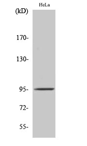
, Peroxidase conjugated at 1/10000 dilution Predicted band size : 95 kDa Blocking/Dilution buffer: 5% NFDM/TBST.")
. The cells were fixed with 2% paraformaldehyde (10 min) and then permeabilized with 90% methanol for 10 min. The cells were then icubated in 2% bovine serum albumin to block non-specific protein-protein interactions followed by the antibody (M01630, 1:25 dilution) for 60 min at 37C. The secondary antibody used was Goat-Anti-Mouse IgG, DyLight® 488 Conjugated Highly Cross-Adsorbed at 1/400 dilution for 40 min at 37C. Isotype control antibody (blue line) was mouse IgG1 (1g/1x10^6 cells) used under the same conditions. Acquisition of >10, 000 events was performed.")
Immunofluorescent analysis of 4% paraformaldehyde-fixed, 0.1% Triton X-100 permeabilized NIH/3T3 (mouse embryonic fibroblast cell line) cells labeling Fer with M01630 at 1/25 dilution, followed by Dylight® 488-conjugated goat anti-mouse IgG secondary antibody at 1/200 dilution (green). Immunofluorescence image showing cytoplasm staining on NIH/3T3 cell line. The nuclear counter stain is DAPI (blue).
Anti-Fer Antibody
M01630
ApplicationsFlow Cytometry, ImmunoFluorescence, Western Blot
Product group Antibodies
TargetFer
Overview
- SupplierBoster Bio
- Product NameAnti-Fer Antibody
- Delivery Days Customer9
- ApplicationsFlow Cytometry, ImmunoFluorescence, Western Blot
- CertificationResearch Use Only
- ClonalityMonoclonal
- Clone ID1487CT794.8.50
- Gene ID14158
- Target nameFer
- Target descriptionFER tyrosine kinase
- Target synonymsC330004K01Rik, Fert, Fert2, tyrosine-protein kinase Fer, fer (fms/fps related) protein kinase, testis specific 2, p94-Fer, proto-oncogene c-Fer, proto-oncogene tyrosine-protein kinase Fer
- HostMouse
- IsotypeIgG2a
- Protein IDP70451
- Protein NameTyrosine-protein kinase Fer
- Scientific DescriptionBoster Bio Anti-Fer Antibody (Catalog # M01630). Tested in WB, Flow Cytometry, IF application(s). This antibody reacts with Mouse.
- Storage Instruction-20°C,2°C to 8°C
- UNSPSC12352203
Related products
Product group Antibodies
Anti-Mouse Fer Antibody102-20135
ApplicationsFlow Cytometry, ImmunoFluorescence, Western Blot
TargetFer
- SizePrice