


Figure 1. Western blot analysis of Ferritin Light Chain using anti-Ferritin Light Chain antibody (M01956-1). Electrophoresis was performed on a 5-20% SDS-PAGE gel at 70V (Stacking gel) / 90V (Resolving gel) for 2-3 hours. The sample well of each lane was loaded with 30 ug of sample under reducing conditions. Lane 1: human HepG2 whole cell lysates, Lane 2: human A549 whole cell lysates, Lane 3: human THP-1 whole cell lysates, Lane 4: human SiHa whole cell lysates, Lane 5: rat liver tissue lysates. After electrophoresis, proteins were transferred to a nitrocellulose membrane at 150 mA for 50-90 minutes. Blocked the membrane with 5% non-fat milk/TBS for 1.5 hour at RT. The membrane was incubated with rabbit anti-Ferritin Light Chain antigen affinity purified monoclonal antibody (Catalog # M01956-1) at 1:1000 overnight at 4°C, then washed with TBS-0.1%Tween 3 times with 5 minutes each and probed with a goat anti-rabbit IgG-HRP secondary antibody at a dilution of 1:500 for 1.5 hour at RT. The signal is developed using an Enhanced Chemiluminescent detection (ECL) kit (Catalog # EK1002) with Tanon 5200 system. A specific band was detected for Ferritin Light Chain at approximately 20 kDa. The expected band size for Ferritin Light Chain is at 20 kDa.
 HepG2 cell lysate; (2) Mouse liver lysate.")
. Ferritin Light Chain was detected in a paraffin-embedded section of human colon tissue. Heat mediated antigen retrieval was performed in EDTA buffer (pH 8.0, epitope retrieval solution). The tissue section was blocked with 10% goat serum. The tissue section was then incubated with 1:50 rabbit anti-Ferritin Light Chain Antibody (M01956-1) overnight at 4°C. Peroxidase Conjugated Goat Anti-rabbit IgG was used as secondary antibody and incubated for 30 minutes at 37°C. The tissue section was developed using HRP Conjugated Rabbit IgG Super Vision Assay Kit (Catalog # SV0002) with DAB as the chromogen.")
. Ferritin Light Chain was detected in a paraffin-embedded section of human colon tissue. Heat mediated antigen retrieval was performed in EDTA buffer (pH 8.0, epitope retrieval solution). The tissue section was blocked with 10% goat serum. The tissue section was then incubated with 1:50 rabbit anti-Ferritin Light Chain Antibody (M01956-1) overnight at 4°C. Peroxidase Conjugated Goat Anti-rabbit IgG was used as secondary antibody and incubated for 30 minutes at 37°C. The tissue section was developed using HRP Conjugated Rabbit IgG Super Vision Assay Kit (Catalog # SV0002) with DAB as the chromogen.")
. Ferritin Light Chain was detected in a paraffin-embedded section of human colon tissue. Heat mediated antigen retrieval was performed in EDTA buffer (pH 8.0, epitope retrieval solution). The tissue section was blocked with 10% goat serum. The tissue section was then incubated with 1:50 rabbit anti-Ferritin Light Chain Antibody (M01956-1) overnight at 4°C. Peroxidase Conjugated Goat Anti-rabbit IgG was used as secondary antibody and incubated for 30 minutes at 37°C. The tissue section was developed using HRP Conjugated Rabbit IgG Super Vision Assay Kit (Catalog # SV0002) with DAB as the chromogen.")
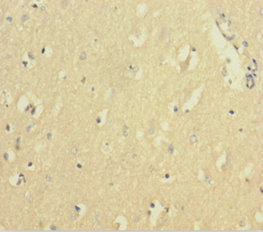
. Ferritin Light Chain was detected in a paraffin-embedded section of human colon tissue. Heat mediated antigen retrieval was performed in EDTA buffer (pH 8.0, epitope retrieval solution). The tissue section was blocked with 10% goat serum. The tissue section was then incubated with 1:50 rabbit anti-Ferritin Light Chain Antibody (M01956-1) overnight at 4°C. Peroxidase Conjugated Goat Anti-rabbit IgG was used as secondary antibody and incubated for 30 minutes at 37°C. The tissue section was developed using HRP Conjugated Rabbit IgG Super Vision Assay Kit (Catalog # SV0002) with DAB as the chromogen.")
Figure 1. Western blot analysis of Ferritin Light Chain using anti-Ferritin Light Chain antibody (M01956-1). Electrophoresis was performed on a 5-20% SDS-PAGE gel at 70V (Stacking gel) / 90V (Resolving gel) for 2-3 hours. The sample well of each lane was loaded with 30 ug of sample under reducing conditions. Lane 1: human HepG2 whole cell lysates, Lane 2: human A549 whole cell lysates, Lane 3: human THP-1 whole cell lysates, Lane 4: human SiHa whole cell lysates, Lane 5: rat liver tissue lysates. After electrophoresis, proteins were transferred to a nitrocellulose membrane at 150 mA for 50-90 minutes. Blocked the membrane with 5% non-fat milk/TBS for 1.5 hour at RT. The membrane was incubated with rabbit anti-Ferritin Light Chain antigen affinity purified monoclonal antibody (Catalog # M01956-1) at 1:1000 overnight at 4°C, then washed with TBS-0.1%Tween 3 times with 5 minutes each and probed with a goat anti-rabbit IgG-HRP secondary antibody at a dilution of 1:500 for 1.5 hour at RT. The signal is developed using an Enhanced Chemiluminescent detection (ECL) kit (Catalog # EK1002) with Tanon 5200 system. A specific band was detected for Ferritin Light Chain at approximately 20 kDa. The expected band size for Ferritin Light Chain is at 20 kDa.
Anti-Ferritin Light Chain FTL Monoclonal Antibody
M01956-1
ApplicationsFlow Cytometry, ImmunoPrecipitation, Western Blot, ImmunoHistoChemistry
Product group Antibodies
ReactivityHuman, Mouse, Rat
TargetFTL
Overview
- SupplierBoster Bio
- Product NameAnti-Ferritin Light Chain FTL Monoclonal Antibody
- Delivery Days Customer9
- ApplicationsFlow Cytometry, ImmunoPrecipitation, Western Blot, ImmunoHistoChemistry
- CertificationResearch Use Only
- ClonalityMonoclonal
- Clone IDACBG-6
- Gene ID2512
- Target nameFTL
- Target descriptionferritin light chain
- Target synonymsFTL1, LFTD, NBIA3, ferritin light chain, epididymis secretory sperm binding protein, ferritin L subunit, ferritin L-chain, ferritin light polypeptide-like 3, ferritin, light polypeptide
- HostRabbit
- IsotypeIgG
- Protein IDP02792
- Protein NameFerritin light chain
- Scientific DescriptionBoster Bio Anti-Ferritin Light Chain FTL Monoclonal Antibody catalog # M01956-1. Tested in WB, IHC, IP, Flow Cytometry applications. This antibody reacts with Human, Mouse, Rat.
- ReactivityHuman, Mouse, Rat
- Storage Instruction-20°C
- UNSPSC12352203
Datasheet
MSDS
Related products

Product group Antibodies
Anti-FTL AntibodyA30603
ApplicationsWestern Blot, ImmunoHistoChemistry
ReactivityHuman, Mouse, Rat
- SizePrice
Product group Antibodies
Anti-FTL Antibody144-01768
ApplicationsImmunoFluorescence, Western Blot, ImmunoHistoChemistry
ReactivityHuman, Mouse
TargetFTL
- SizePrice

Product group Antibodies

FTL / Ferritin Light Chain AntibodyLS-C834943
ApplicationsELISA, ImmunoHistoChemistry
ReactivityHuman
TargetFTL
- SizePrice

Product group Antibodies
ApplicationsFlow Cytometry, ImmunoPrecipitation, Western Blot
ReactivityHuman, Mouse, Rat
TargetFTL
- SizePrice
Product group Antibodies
FTL AntibodyCSB-PA11407A0RB
ApplicationsELISA, ImmunoHistoChemistry
ReactivityHuman
TargetFTL
- SizePrice
Product group Antibodies

Goat anti-FTL, BiotinylatedEB09092-B
ApplicationsWestern Blot, ELISA, ImmunoHistoChemistry
ReactivityBovine, Canine, Human, Mouse, Rat
TargetFTL
- SizePrice
Product group Antibodies
Ftl Polyclonal AntibodyCAC09142
ApplicationsImmunoFluorescence, ELISA, ImmunoHistoChemistry
TargetFTL
- SizePrice
![Mouse tissue extract (50 μg) was separated by 12% SDS-PAGE, and the membrane was blotted with Ferritin Light Chain antibody [N1C3] (GTX101211) diluted at 1:1000.](https://www.genetex.com/upload/website/prouct_img/normal/GTX101211/GTX101211_39694_20160714_WB_M_liver_w_23060100_787.webp)
Product group Antibodies
Ferritin Light Chain antibody [N1C3]GTX101211
ApplicationsWestern Blot, ImmunoHistoChemistry, ImmunoHistoChemistry Paraffin
ReactivityHuman, Mouse
TargetFTL
- SizePrice