


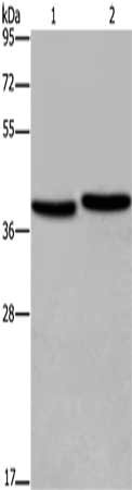
Figure 1. Western blot analysis of FHL1 using anti-FHL1 antibody (A01258-3). Electrophoresis was performed on a 5-20% SDS-PAGE gel at 70V (Stacking gel) / 90V (Resolving gel) for 2-3 hours. The sample well of each lane was loaded with 30 ug of sample under reducing conditions. Lane 1: rat heart tissue lysates, Lane 2: mouse heart tissue lysates. After electrophoresis, proteins were transferred to a nitrocellulose membrane at 150 mA for 50-90 minutes. Blocked the membrane with 5% non-fat milk/TBS for 1.5 hour at RT. The membrane was incubated with rabbit anti-FHL1 antigen affinity purified polyclonal antibody (Catalog # A01258-3) at 0.5 microg/mL overnight at 4°C, then washed with TBS-0.1%Tween 3 times with 5 minutes each and probed with a goat anti-rabbit IgG-HRP secondary antibody at a dilution of 1:5000 for 1.5 hour at RT. The signal is developed using an Enhanced Chemiluminescent detection (ECL) kit (Catalog # EK1002) with Tanon 5200 system. A specific band was detected for FHL1 at approximately 36 kDa. The expected band size for FHL1 is at 36 kDa.
Figure 1. Western blot analysis of FHL1 using anti-FHL1 antibody (A01258-3). Electrophoresis was performed on a 5-20% SDS-PAGE gel at 70V (Stacking gel) / 90V (Resolving gel) for 2-3 hours. The sample well of each lane was loaded with 30 ug of sample under reducing conditions. Lane 1: rat heart tissue lysates, Lane 2: mouse heart tissue lysates. After electrophoresis, proteins were transferred to a nitrocellulose membrane at 150 mA for 50-90 minutes. Blocked the membrane with 5% non-fat milk/TBS for 1.5 hour at RT. The membrane was incubated with rabbit anti-FHL1 antigen affinity purified polyclonal antibody (Catalog # A01258-3) at 0.5 microg/mL overnight at 4°C, then washed with TBS-0.1%Tween 3 times with 5 minutes each and probed with a goat anti-rabbit IgG-HRP secondary antibody at a dilution of 1:5000 for 1.5 hour at RT. The signal is developed using an Enhanced Chemiluminescent detection (ECL) kit (Catalog # EK1002) with Tanon 5200 system. A specific band was detected for FHL1 at approximately 36 kDa. The expected band size for FHL1 is at 36 kDa.
Anti-FHL1 Antibody Picoband(r)
A01258-3-FITC
ApplicationsWestern Blot, ELISA
Product group Antibodies
ReactivityHuman, Mouse, Rat
TargetFHL1
Overview
- SupplierBoster Bio
- Product NameAnti-FHL1 Antibody Picoband(r)
- Delivery Days Customer9
- ApplicationsWestern Blot, ELISA
- CertificationResearch Use Only
- ClonalityPolyclonal
- Concentration500 ug/ml
- ConjugateFITC
- Gene ID2273
- Target nameFHL1
- Target descriptionfour and a half LIM domains 1
- Target synonymsFCMSU, FHL-1, FHL1A, FHL1B, FLH1A, KYOT, RBMX1A, RBMX1B, SLIM, SLIM-1, SLIM1, SLIMMER, XMPMA, four and a half LIM domains protein 1, LIM protein SLIMMER, four-and-a-half Lin11, Isl-1 and Mec-3 domains 1, skeletal muscle LIM-protein 1
- HostRabbit
- IsotypeIgG
- Protein IDQ13642
- Protein NameFour and a half LIM domains protein 1
- Scientific DescriptionBoster Bio Anti-FHL1 Antibody Picoband® catalog # A01258-3. Tested in ELISA, WB applications. This antibody reacts with Human, Mouse, Rat. The brand Picoband indicates this is a premium antibody that guarantees superior quality, high affinity, and strong signals with minimal background in Western blot applications. Only our best-performing antibodies are designated as Picoband, ensuring unmatched performance.
- ReactivityHuman, Mouse, Rat
- Storage Instruction-20°C,2°C to 8°C
- UNSPSC12352203
Related products
Product group Antibodies
FHL1 AntibodyCSB-PA174115
ApplicationsWestern Blot, ELISA
ReactivityHuman, Mouse, Rat
TargetFHL1
- SizePrice
Product group Antibodies
Anti-FHL1 AntibodyA14777
ApplicationsWestern Blot
ReactivityHuman, Mouse
- SizePrice

Product group Antibodies
Anti-FHL FHL1 Antibody Picoband(r)A01258-1-CARRIER-FREE
ApplicationsFlow Cytometry, ImmunoFluorescence, Western Blot, ELISA, ImmunoCytoChemistry, ImmunoHistoChemistry
ReactivityHuman
TargetFHL1
- SizePrice
Product group Antibodies
References

ApplicationsWestern Blot, ELISA
ReactivityBovine, Canine, Human, Mouse, Porcine, Rat
TargetFHL1
- SizePrice

Product group Antibodies
Anti-FHL1 AntibodyHPA001040
ApplicationsWestern Blot, ImmunoCytoChemistry, ImmunoHistoChemistry
ReactivityHuman
TargetFHL1
- SizePrice
Product group Antibodies

SLIM / FHL1 AntibodyLS-C401682
ApplicationsWestern Blot, ELISA
ReactivityHuman, Mouse, Rat
TargetFHL1
- SizePrice
Product group Antibodies
FHL1 Recombinant Antibody, AbBy Fluor-488 ConjugatedBSM-61727R-BF488
ApplicationsWestern Blot
ReactivityHuman, Mouse, Rat
TargetFHL1
- SizePrice
Product group Antibodies
Fhl1 Polyclonal AntibodyCAC08171
ApplicationsImmunoFluorescence, Western Blot, ELISA
ReactivityMouse, Rat
TargetFHL1
- SizePrice
Product group Antibodies
FHL1 antibodyGTX100266
ApplicationsWestern Blot
ReactivityHuman, Mouse
TargetFHL1
- SizePrice