


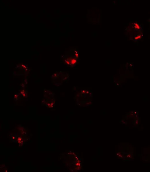
Immunohistochemical staining of human kidney shows strong cytoplasmic positivity in renal tubules.
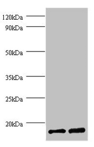
![Lane 1: Marker [kDa] 250, 130, 100, 70, 55, 35, 25, 15, 10. Lane 2: Human cell line HepG2](https://atlasantibodies.s3.amazonaws.com/images/wb/hpa051798-wb-1.jpg "Lane 1: Marker [kDa] 250, 130, 100, 70, 55, 35, 25, 15, 10. Lane 2: Human cell line HepG2")
Immunohistochemical staining of human kidney shows strong cytoplasmic positivity in renal tubules.
Anti-FKBP1A Antibody
HPA051798
ApplicationsWestern Blot, ImmunoHistoChemistry
Product group Antibodies
ReactivityHuman
TargetFKBP1A
Overview
- SupplierAtlas Antibodies
- Product NameAnti-FKBP1A Antibody
- Delivery Days Customer4
- ApplicationsWestern Blot, ImmunoHistoChemistry
- CertificationResearch Use Only
- ClonalityPolyclonal
- ConjugateUnconjugated
- Gene ID2280
- Target nameFKBP1A
- Target descriptionFKBP prolyl isomerase 1A
- Target synonymsFKBP-12, FKBP-1A, FKBP1, FKBP12, PKC12, PKCI2, PPIASE, peptidyl-prolyl cis-trans isomerase FKBP1A, 12 kDa FK506-binding protein, 12 kDa FKBP, FK506 binding protein 1A, 12kDa, FK506 binding protein12, FK506-binding protein 1, FK506-binding protein 12, FK506-binding protein 1A, FK506-binding protein, T-cell, 12-kD, FKBP12-Exip3, PPIase FKBP1A, calstabin-1, immunophilin FKBP12, protein kinase C inhibitor 2, rotamase
- HostRabbit
- IsotypeIgG
- Protein IDP62942
- Protein NamePeptidyl-prolyl cis-trans isomerase FKBP1A
- Scientific DescriptionRecombinant Protein Epitope Signature Tag (PrEST) antigen sequence
- ReactivityHuman
- Storage Instruction-20°C,2°C to 8°C
- UNSPSC41116161
Datasheet
MSDS
Related products
Product group Antibodies
FKBP1A AntibodyCSB-PA02325A0RB
ApplicationsImmunoFluorescence, Western Blot, ELISA, ImmunoHistoChemistry
ReactivityHuman
TargetFKBP1A
- SizePrice
Product group Antibodies
FKBP1A Polyclonal AntibodyCAC13952
ApplicationsImmunoFluorescence, Western Blot, ELISA, ImmunoHistoChemistry
TargetFKBP1A
- SizePrice
Product group Antibodies
Anti-FKBP1A Antibody144-01763
ApplicationsWestern Blot, ImmunoHistoChemistry
ReactivityHuman, Mouse, Rat
TargetFKBP1A
- SizePrice

Product group Antibodies
Anti-FKBP1A AntibodyA29863
ApplicationsWestern Blot, ImmunoHistoChemistry
ReactivityHuman, Mouse, Rat
- SizePrice
Product group Antibodies

FKBP1A / FKBP12 AntibodyLS-C832161
ApplicationsELISA, ImmunoHistoChemistry
ReactivityHuman, Mouse, Rat
TargetFKBP1A
- SizePrice

Product group Antibodies
Anti-FKBP1A/1B Antibody Picoband(r)A04492-3-CARRIER-FREE
ApplicationsFlow Cytometry, Western Blot, ELISA, ImmunoHistoChemistry
ReactivityHuman, Mouse
TargetFKBP1A
- SizePrice
Product group Antibodies
FKBP12 antibodyGTX31576
ApplicationsImmunoFluorescence, Western Blot, ELISA, ImmunoCytoChemistry
ReactivityHuman, Mouse, Rat
TargetFKBP1A
- SizePrice