


Immunohistochemical staining of human testis shows moderate to strong cytoplasmic positivity in cells in seminiferous ducts.

Immunohistochemical staining of human testis shows moderate to strong cytoplasmic positivity in cells in seminiferous ducts.
Anti-FXR1-25ul
HPA018246
ApplicationsWestern Blot, ImmunoCytoChemistry, ImmunoHistoChemistry
Product group Antibodies
ReactivityHuman, Mouse, Rat
Overview
- SupplierAtlas Antibodies
- Product NameAnti-FXR1-25ul
- Delivery Days Customer6
- ApplicationsWestern Blot, ImmunoCytoChemistry, ImmunoHistoChemistry
- Applications SupplierICC, WB, IHC
- CertificationResearch Use Only
- ClonalityPolyclonal
- Concentration0.1
- ConjugateUnconjugated
- HostRabbit
- IsotypeIgG
- Protein IDP51114
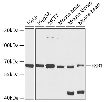
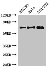
- Protein NameRNA-binding protein FXR1
- Scientific DescriptionRabbit Polyclonal Anti-FXR1 Antibody against Human fragile X mental retardation, autosomal homolog 1. Validated for Immunofluorescence, Immunohistochemistry and Western Blot
- ReactivityHuman, Mouse, Rat
- Storage InstructionStore at +4°C for short term storage. Long time storage is recommended at -20°C.
- UNSPSC12352203
Datasheet
MSDS
Related products
Product group Antibodies
Anti-FXR1 AntibodyA15059
ApplicationsWestern Blot
ReactivityHuman, Mouse, Rat
- SizePrice

Product group Antibodies
Anti-FXR1 Antibody Picoband(r)A03308-2-CARRIER-FREE
ApplicationsFlow Cytometry, ImmunoFluorescence, Western Blot, ELISA, ImmunoCytoChemistry, ImmunoHistoChemistry
ReactivityHuman, Monkey, Mouse, Rat
TargetFXR1
- SizePrice
Product group Antibodies
Anti-FXR1 Antibody144-05942
ApplicationsImmunoFluorescence, Western Blot
ReactivityHuman, Mouse, Rat
TargetFXR1
- SizePrice
Product group Antibodies
FXR1 Recombinant AntibodyBSM-62314R
ApplicationsFlow Cytometry, ImmunoFluorescence, Western Blot, ImmunoCytoChemistry, ImmunoHistoChemistry, ImmunoHistoChemistry Frozen, ImmunoHistoChemistry Paraffin
ReactivityHuman, Mouse, Rat
TargetFXR1
- SizePrice
Product group Antibodies
FXR1 AntibodyCSB-PA009087LA01HU
ApplicationsImmunoFluorescence, Western Blot, ELISA, ImmunoHistoChemistry
ReactivityHuman, Mouse
TargetFXR1
- SizePrice
Product group Antibodies

Goat anti-FXR1EB07361
ApplicationsFlow Cytometry, ImmunoFluorescence, Western Blot, ELISA
ReactivityBovine, Canine, Human, Mouse, Rat
TargetFXR1
- SizePrice
Product group Antibodies
FXR1 Polyclonal AntibodyCAC14724
ApplicationsImmunoFluorescence, Western Blot, ELISA, ImmunoHistoChemistry
ReactivityMouse
TargetFXR1
- SizePrice
Product group Antibodies

FXR1 AntibodyLS-C334404
ApplicationsWestern Blot
ReactivityHuman, Mouse, Rat
TargetFXR1
- SizePrice