

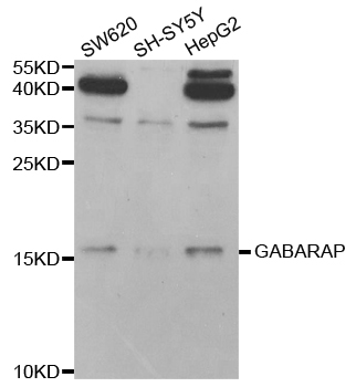
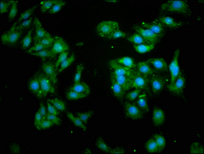
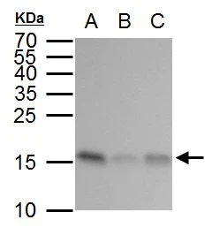
Anti-GABARAP Antibody
144-12568
ApplicationsWestern Blot, ImmunoHistoChemistry
Product group Antibodies
ReactivityHuman, Mouse, Rat
TargetGABARAP
Overview
- SupplierRayBiotech
- Product NameAnti-GABARAP Antibody
- Delivery Days Customer16
- ApplicationsWestern Blot, ImmunoHistoChemistry
- CertificationResearch Use Only
- ClonalityPolyclonal
- ConjugateUnconjugated
- Gene ID11337
- Target nameGABARAP
- Target descriptionGABA type A receptor-associated protein
- Target synonymsATG8A, GABARAP-a, MM46, gamma-aminobutyric acid receptor-associated protein, GABA(A) receptor-associated protein, epididymis secretory sperm binding protein
- HostRabbit
- IsotypeIgG
- Protein IDO95166
- Protein NameGamma-aminobutyric acid receptor-associated protein
- Scientific DescriptionGABARAP Polyclonal Antibody
- ReactivityHuman, Mouse, Rat
- Storage Instruction-20°C
- UNSPSC12352203
Related products
Product group Antibodies
Anti-GABARAP AntibodyA30839
ApplicationsImmunoFluorescence, Western Blot, ImmunoHistoChemistry
ReactivityHuman, Mouse, Rat
- SizePrice
Product group Antibodies

GABARAP AntibodyLS-C747664
ApplicationsWestern Blot, ImmunoHistoChemistry
ReactivityHuman, Mouse, Rat
TargetGABARAP
- SizePrice

Product group Antibodies
Anti-GABARAP Antibody Picoband(r)A01907-2-CARRIER-FREE
ApplicationsFlow Cytometry, Western Blot, ImmunoHistoChemistry
ReactivityHuman, Mouse, Rat
TargetGABARAP
- SizePrice
Product group Antibodies
GABARAP Recombinant AntibodyBSM-61023R
ApplicationsImmunoFluorescence, Western Blot, ImmunoHistoChemistry, ImmunoHistoChemistry Frozen, ImmunoHistoChemistry Paraffin
ReactivityHuman, Mouse, Rat
TargetGABARAP
- SizePrice
Product group Antibodies
Gabarap Polyclonal AntibodyCAC07105
ApplicationsImmunoFluorescence, ELISA
TargetGABARAP
- SizePrice
Product group Antibodies
GABARAP AntibodyCSB-PA05484A0RB
ApplicationsImmunoFluorescence, ELISA
ReactivityHuman
TargetGABARAP
- SizePrice
Product group Antibodies
GABARAP antibodyGTX129710
ApplicationsImmunoFluorescence, Western Blot, ImmunoCytoChemistry
ReactivityHuman
TargetGABARAP
- SizePrice
Product group Antibodies
ApplicationsWestern Blot, ELISA
ReactivityHuman
TargetGABARAP
- SizePrice