


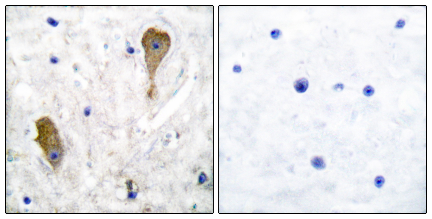
Immunohistochemical staining of rat hippocampal formation shows strong positivity in GABAergic fibers in the CA1 area.
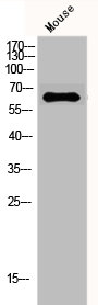
![Lane 1: Marker [kDa] 250, 130, 100, 70, 55, 35, 25, 15, 10. Lane 2: Human Cerebral Cortex tissue](https://atlasantibodies.s3.amazonaws.com/images/wb/amab91079-wb-1.jpg "Lane 1: Marker [kDa] 250, 130, 100, 70, 55, 35, 25, 15, 10. Lane 2: Human Cerebral Cortex tissue")
Immunohistochemical staining of rat hippocampal formation shows strong positivity in GABAergic fibers in the CA1 area.
Anti-GAD1 Antibody
AMAB91079
ApplicationsWestern Blot, ImmunoHistoChemistry
Product group Antibodies
ReactivityHuman, Mouse, Rat
TargetGAD1
Overview
- SupplierAtlas Antibodies
- Product NameAnti-GAD1 Antibody
- Delivery Days Customer4
- ApplicationsWestern Blot, ImmunoHistoChemistry
- CertificationResearch Use Only
- ClonalityMonoclonal
- Clone IDCL2919
- ConjugateUnconjugated
- Gene ID2571
- Target nameGAD1
- Target descriptionglutamate decarboxylase 1
- Target synonymsCPSQ1, DEE89, GAD, SCP, glutamate decarboxylase 1, 67 kDa glutamic acid decarboxylase, GAD-67, glutamate decarboxylase 1 (brain, 67kDa)
- HostMouse
- IsotypeIgG2b
- Protein IDQ99259
- Protein NameGlutamate decarboxylase 1
- Scientific DescriptionRecombinant Protein Epitope Signature Tag (PrEST) antigen sequence
- ReactivityHuman, Mouse, Rat
- Storage Instruction-20°C,2°C to 8°C
- UNSPSC41116161
Datasheet
MSDS
Related products
Product group Antibodies
Anti-GAD1 AntibodyA96199
ApplicationsWestern Blot, ELISA, ImmunoHistoChemistry
ReactivityHuman, Mouse, Rat
- SizePrice
Product group Antibodies
Anti-GAD1 Antibody144-02938
ApplicationsWestern Blot
ReactivityHuman, Mouse, Rat
TargetGAD1
- SizePrice

Product group Antibodies
Anti-GAD1 AntibodyAMAB91076
ApplicationsWestern Blot, ImmunoHistoChemistry
ReactivityHuman, Mouse, Rat
TargetGAD1
- SizePrice
Product group Antibodies
Anti-GAD1 AntibodyAMAB91076
ApplicationsWestern Blot, ImmunoHistoChemistry
ReactivityHuman, Mouse, Rat
TargetGAD1
- SizePrice

Product group Antibodies
Anti-GAD1 AntibodyAMAB91078
ApplicationsWestern Blot, ImmunoHistoChemistry
ReactivityHuman, Mouse, Rat
TargetGAD1
- SizePrice
Product group Antibodies
References
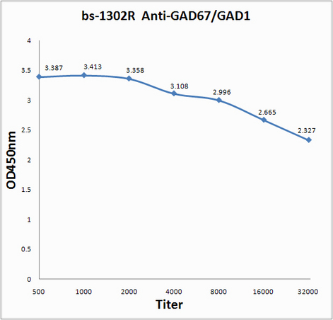
GAD67 Polyclonal AntibodyBS-1302R
ApplicationsImmunoFluorescence, Western Blot, ELISA, ImmunoCytoChemistry, ImmunoHistoChemistry, ImmunoHistoChemistry Frozen, ImmunoHistoChemistry Paraffin
ReactivityHuman, Mouse, Rat
TargetGAD1
- SizePrice
Product group Antibodies
GAD1/GAD2 AntibodyCSB-PA002622
ApplicationsImmunoFluorescence, Western Blot, ELISA, ImmunoHistoChemistry
ReactivityHuman, Mouse
TargetGAD1
- SizePrice