


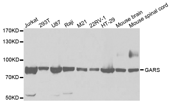
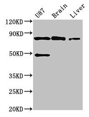
Figure 1. Western blot analysis of GARS1 using anti-GARS1 antibody (A04618-2). Electrophoresis was performed on a 5-20% SDS-PAGE gel at 70V (Stacking gel) / 90V (Resolving gel) for 2-3 hours. The sample well of each lane was loaded with 30 ug of sample under reducing conditions. Lane 1: human HT-1080 whole cell lysates, Lane 2: human HepG2 whole cell lysates, Lane 3: human Hela whole cell lysates, Lane 4: human Jurkat whole cell lysates, Lane 5: rat brain tissue lysates, Lane 6: rat RH35 whole cell lysates, Lane 7: mouse brain tissue lysates, Lane 8: mouse Neuro-2a whole cell lysates. After electrophoresis, proteins were transferred to a nitrocellulose membrane at 150 mA for 50-90 minutes. Blocked the membrane with 5% non-fat milk/TBS for 1.5 hour at RT. The membrane was incubated with rabbit anti-GARS1 antigen affinity purified polyclonal antibody (Catalog # A04618-2) at 0.5 microg/mL overnight at 4°C, then washed with TBS-0.1%Tween 3 times with 5 minutes each and probed with a goat anti-rabbit IgG-HRP secondary antibody at a dilution of 1:5000 for 1.5 hour at RT. The signal is developed using an Enhanced Chemiluminescent detection (ECL) kit (Catalog # EK1002) with Tanon 5200 system. A specific band was detected for GARS1 at approximately 80 kDa. The expected band size for GARS1 is at 83 kDa.
. Overlay histogram showing HepG2 cells stained with A04618-2 (Blue line). The cells were fixed with 4% paraformaldehyde and blocked with 10% normal goat serum. And then incubated with rabbit anti-GARS1 Antibody (A04618-2, 1 microg/1x106 cells) for 30 min at 20°C. DyLight®488 conjugated goat anti-rabbit IgG (BA1127, 5-10 microg/1x106 cells) was used as secondary antibody for 30 minutes at 20°C. Isotype control antibody (Green line) was rabbit IgG (1 microg/1x106) used under the same conditions. Unlabelled sample (Red line) was also used as a control.")
Figure 1. Western blot analysis of GARS1 using anti-GARS1 antibody (A04618-2). Electrophoresis was performed on a 5-20% SDS-PAGE gel at 70V (Stacking gel) / 90V (Resolving gel) for 2-3 hours. The sample well of each lane was loaded with 30 ug of sample under reducing conditions. Lane 1: human HT-1080 whole cell lysates, Lane 2: human HepG2 whole cell lysates, Lane 3: human Hela whole cell lysates, Lane 4: human Jurkat whole cell lysates, Lane 5: rat brain tissue lysates, Lane 6: rat RH35 whole cell lysates, Lane 7: mouse brain tissue lysates, Lane 8: mouse Neuro-2a whole cell lysates. After electrophoresis, proteins were transferred to a nitrocellulose membrane at 150 mA for 50-90 minutes. Blocked the membrane with 5% non-fat milk/TBS for 1.5 hour at RT. The membrane was incubated with rabbit anti-GARS1 antigen affinity purified polyclonal antibody (Catalog # A04618-2) at 0.5 microg/mL overnight at 4°C, then washed with TBS-0.1%Tween 3 times with 5 minutes each and probed with a goat anti-rabbit IgG-HRP secondary antibody at a dilution of 1:5000 for 1.5 hour at RT. The signal is developed using an Enhanced Chemiluminescent detection (ECL) kit (Catalog # EK1002) with Tanon 5200 system. A specific band was detected for GARS1 at approximately 80 kDa. The expected band size for GARS1 is at 83 kDa.
Anti-GARS1 Antibody Picoband(r)
A04618-2-CARRIER-FREE
ApplicationsFlow Cytometry, Western Blot, ELISA
Product group Antibodies
ReactivityHuman, Mouse, Rat
TargetGARS1
Overview
- SupplierBoster Bio
- Product NameAnti-GARS1 Antibody Picoband(r)
- Delivery Days Customer9
- ApplicationsFlow Cytometry, Western Blot, ELISA
- CertificationResearch Use Only
- ClonalityPolyclonal
- Concentration500 ug/ml
- Gene ID2617
- Target nameGARS1
- Target descriptionglycyl-tRNA synthetase 1
- Target synonymsCMT2D, DSMAV, GARS, GlyRS, HMN5, HMN5A, HMND5, SMAD1, SMAJI, glycine--tRNA ligase, AP-4-A synthetase, Charcot-Marie-Tooth neuropathy 2D, Charcot-Marie-Tooth neuropathy, neuronal type, D, ap4A synthetase, diadenosine tetraphosphate synthetase
- HostRabbit
- Protein IDP41250
- Protein NameGlycine--tRNA ligase
- Scientific DescriptionBoster Bio Anti-GARS1 Antibody Picoband® catalog # A04618-2. Tested in WB, Flow Cytometry, ELISA applications. This antibody reacts with Human, Mouse, Rat. The brand Picoband indicates this is a premium antibody that guarantees superior quality, high affinity, and strong signals with minimal background in Western blot applications. Only our best-performing antibodies are designated as Picoband, ensuring unmatched performance.
- ReactivityHuman, Mouse, Rat
- Storage Instruction-20°C,2°C to 8°C
- UNSPSC12352203
Related products
Product group Antibodies
Anti-GARS AntibodyA30783
ApplicationsImmunoFluorescence, Western Blot, ImmunoHistoChemistry
ReactivityHuman, Mouse, Rat
- SizePrice
Product group Antibodies
Anti-GARS Antibody144-60275
ApplicationsWestern Blot
ReactivityHuman, Mouse
TargetGARS1
- SizePrice
Product group Antibodies

GARS / Glycyl tRNA Synthetase AntibodyLS-C830460
ApplicationsWestern Blot, ELISA
ReactivityHuman, Mouse, Rat
TargetGARS1
- SizePrice
Product group Antibodies
ApplicationsWestern Blot, ImmunoCytoChemistry
ReactivityHuman, Mouse, Rat
TargetGARS1
- SizePrice
Product group Antibodies
GARS AntibodyCSB-PA009262EA01HU
ApplicationsImmunoFluorescence, ImmunoPrecipitation, Western Blot, ELISA, ImmunoHistoChemistry
ReactivityHuman, Mouse, Rat
TargetGARS1
- SizePrice
Product group Antibodies
GARS Polyclonal AntibodyCAC14725
ApplicationsImmunoFluorescence, ImmunoPrecipitation, Western Blot, ELISA, ImmunoHistoChemistry
ReactivityMouse, Rat
TargetGARS1
- SizePrice
![GARS antibody [N3C2], Internal detects GARS protein at cytosol on human colon carcinoma by immunohistochemical analysis. Sample: Paraffin-embedded colon carcinoma. GARS antibody [N3C2], Internal (GTX114421) dilution: 1:500.
Antigen Retrieval: Trilogy? (EDTA based, pH 8.0) buffer, 15min](https://www.genetex.com/upload/website/prouct_img/normal/GTX114421/GTX114421_40184_IHC_w_23060501_891.webp)
Product group Antibodies
GARS antibody [N3C2], InternalGTX114421
ApplicationsWestern Blot, ImmunoHistoChemistry, ImmunoHistoChemistry Paraffin
ReactivityHuman
TargetGARS1
- SizePrice

Product group Antibodies
Anti-GARS AntibodyHPA019097
ApplicationsWestern Blot, ImmunoHistoChemistry
ReactivityHuman, Mouse, Rat
TargetGARS1
- SizePrice