


Immunofluorescent staining of human cell line HEK 293 shows localization to nuclear speckles.
Immunofluorescent staining of human cell line HEK 293 shows localization to nuclear speckles.
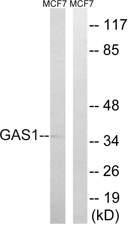
Anti-GAS1 Antibody
HPA036085
ApplicationsImmunoCytoChemistry
Product group Antibodies
ReactivityHuman
TargetGAS1
Overview
- SupplierAtlas Antibodies
- Product NameAnti-GAS1 Antibody
- Delivery Days Customer4
- ApplicationsImmunoCytoChemistry
- CertificationResearch Use Only
- ClonalityPolyclonal
- ConjugateUnconjugated
- Gene ID2619
- Target nameGAS1
- Target descriptiongrowth arrest specific 1
- Target synonymsgrowth arrest-specific protein 1, GAS-1, Growth arrest-specific gene-1
- HostRabbit
- IsotypeIgG
- Protein IDP54826
- Protein NameGrowth arrest-specific protein 1
- Scientific DescriptionRecombinant Protein Epitope Signature Tag (PrEST) antigen sequence
- ReactivityHuman
- Storage Instruction-20°C,2°C to 8°C
- UNSPSC41116161
Datasheet
MSDS
Related products
Product group Antibodies
Gas1 Polyclonal AntibodyCAC11192
ApplicationsImmunoFluorescence, Western Blot, ELISA, ImmunoHistoChemistry
TargetGAS1
- SizePrice
Product group Antibodies
Anti-GAS1 (N-term) Antibody102-20957
ApplicationsFlow Cytometry, ImmunoFluorescence, Western Blot, ImmunoHistoChemistry, ImmunoHistoChemistry Paraffin
TargetGAS1
- SizePrice
Product group Antibodies
Anti-GAS1 AntibodyA99409
ApplicationsWestern Blot, ELISA
ReactivityHuman, Mouse
- SizePrice
Product group Antibodies
References
GAS1 antibodyGTX101732
ApplicationsWestern Blot
ReactivityHuman
TargetGAS1
- SizePrice

Product group Antibodies
Gas1 Polyclonal AntibodyBS-6385R
ApplicationsImmunoFluorescence, Western Blot, ELISA, ImmunoCytoChemistry, ImmunoHistoChemistry, ImmunoHistoChemistry Frozen, ImmunoHistoChemistry Paraffin
ReactivityBovine, Canine, Chicken, Human, Mouse, Porcine, Rat, Sheep
TargetGAS1
- SizePrice
Product group Antibodies

GAS1 AntibodyLS-C670265
ApplicationsWestern Blot, ELISA, ImmunoHistoChemistry, ImmunoHistoChemistry Paraffin
ReactivityHuman
TargetGAS1
- SizePrice

Product group Antibodies
Anti-GAS1 AntibodyHPA066902
ApplicationsImmunoHistoChemistry
ReactivityHuman
TargetGAS1
- SizePrice
Product group Antibodies
GAS1 AntibodyCSB-PA008695
ApplicationsWestern Blot, ELISA
ReactivityHuman, Mouse
TargetGAS1
- SizePrice

Product group Antibodies
Anti-GAS1 Antibody Picoband(r)A06815-2-CARRIER-FREE
ApplicationsFlow Cytometry, Western Blot, ELISA
ReactivityHuman
TargetGAS1
- SizePrice