


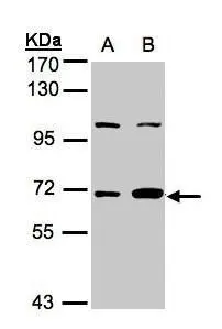
Figure 1. Western blot analysis of GBP1 using anti-GBP1 antibody (A03067-1). Electrophoresis was performed on a 5-20% SDS-PAGE gel at 70V (Stacking gel) / 90V (Resolving gel) for 2-3 hours. The sample well of each lane was loaded with 30 ug of sample under reducing conditions. Lane 1: human placenta tissue lysates, Lane 2: human HUVEC whole cell lysates, Lane 3: human MCF-7 whole cell lysates, Lane 4: human Hela whole cell lysates, Lane 5: rat brain tissue lysates, Lane 6: rat spleen tissue lysates, Lane 7: mouse brain tissue lysates, Lane 8: mouse spleen tissue lysayes. After electrophoresis, proteins were transferred to a nitrocellulose membrane at 150 mA for 50-90 minutes. Blocked the membrane with 5% non-fat milk/TBS for 1.5 hour at RT. The membrane was incubated with rabbit anti-GBP1 antigen affinity purified polyclonal antibody (Catalog # A03067-1) at 0.5 microg/mL overnight at 4°C, then washed with TBS-0.1%Tween 3 times with 5 minutes each and probed with a goat anti-rabbit IgG-HRP secondary antibody at a dilution of 1:5000 for 1.5 hour at RT. The signal is developed using an Enhanced Chemiluminescent detection (ECL) kit (Catalog # EK1002) with Tanon 5200 system. A specific band was detected for GBP1 at approximately 68 kDa. The expected band size for GBP1 is at 68 kDa.
. GBP1 was detected in a paraffin-embedded section of human ovarian cancer tissue. Heat mediated antigen retrieval was performed in EDTA buffer (pH 8.0, epitope retrieval solution). The tissue section was blocked with 10% goat serum. The tissue section was then incubated with 2 microg/ml rabbit anti-GBP1 Antibody (A03067-1) overnight at 4°C. Peroxidase Conjugated Goat Anti-rabbit IgG was used as secondary antibody and incubated for 30 minutes at 37°C. The tissue section was developed using HRP Conjugated Rabbit IgG Super Vision Assay Kit (Catalog # SV0002) with DAB as the chromogen.")
. GBP1 was detected in a paraffin-embedded section of mouse heart tissue. Heat mediated antigen retrieval was performed in EDTA buffer (pH 8.0, epitope retrieval solution). The tissue section was blocked with 10% goat serum. The tissue section was then incubated with 2 microg/ml rabbit anti-GBP1 Antibody (A03067-1) overnight at 4°C. Peroxidase Conjugated Goat Anti-rabbit IgG was used as secondary antibody and incubated for 30 minutes at 37°C. The tissue section was developed using HRP Conjugated Rabbit IgG Super Vision Assay Kit (Catalog # SV0002) with DAB as the chromogen.")
. GBP1 was detected in a paraffin-embedded section of rat heart tissue. Heat mediated antigen retrieval was performed in EDTA buffer (pH 8.0, epitope retrieval solution). The tissue section was blocked with 10% goat serum. The tissue section was then incubated with 2 microg/ml rabbit anti-GBP1 Antibody (A03067-1) overnight at 4°C. Peroxidase Conjugated Goat Anti-rabbit IgG was used as secondary antibody and incubated for 30 minutes at 37°C. The tissue section was developed using HRP Conjugated Rabbit IgG Super Vision Assay Kit (Catalog # SV0002) with DAB as the chromogen.")
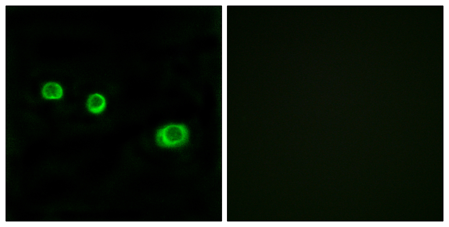
. GBP1 was detected in an immunocytochemical section of SiHa cells. Enzyme antigen retrieval was performed using IHC enzyme antigen retrieval reagent (AR0022) for 15 mins. The cells were blocked with 10% goat serum. And then incubated with 5 microg/mL rabbit anti-GBP1 Antibody (A03067-1) overnight at 4°C. DyLight®488 Conjugated Goat Anti-Rabbit IgG (BA1127) was used as secondary antibody at 1:500 dilution and incubated for 30 minutes at 37°C. The section was counterstained with DAPI. Visualize using a fluorescence microscope and filter sets appropriate for the label used.")
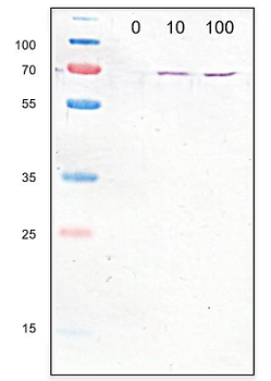
. Lane 1: Hela whole cell lysates (30ug) Lane 2: Rabbit control IgG instead of anti-GBP1 antibody in Hela whole cell lysate. Lane 3: anti-GBP1 antibody (2microg) + Hela whole cell lysate (500microg) After electrophoresis, proteins were transferred to a membrane. Then the membrane was incubated with rabbit anti-GBP1 antigen affinity purified polyclonal antibody (A03067-1) at a dilution of 0.5 microg/mL and probed with a goat anti-rabbit IgG-HRP secondary antibody (Catalog # BA1054). The signal is developed using ECL Plus Western Blotting Substrate (Catalog # AR1197). A specific band was detected for GBP1 at approximately 68 kDa. The expected band size for GBP1 is at 68 kDa.")
. Overlay histogram showing MCF-7 cells stained with A03067-1 (Blue line). The cells were fixed with 4% paraformaldehyde and blocked with 10% normal goat serum. And then incubated with rabbit anti-GBP1 Antibody (A03067-1, 1 microg/1x106 cells) for 30 min at 20°C. DyLight®488 conjugated goat anti-rabbit IgG (BA1127, 5-10 microg/1x106 cells) was used as secondary antibody for 30 minutes at 20°C. Isotype control antibody (Green line) was rabbit IgG (1 microg/1x106) used under the same conditions. Unlabelled sample without incubation with primary antibody and secondary antibody (Red line) was used as a blank control.")
Figure 1. Western blot analysis of GBP1 using anti-GBP1 antibody (A03067-1). Electrophoresis was performed on a 5-20% SDS-PAGE gel at 70V (Stacking gel) / 90V (Resolving gel) for 2-3 hours. The sample well of each lane was loaded with 30 ug of sample under reducing conditions. Lane 1: human placenta tissue lysates, Lane 2: human HUVEC whole cell lysates, Lane 3: human MCF-7 whole cell lysates, Lane 4: human Hela whole cell lysates, Lane 5: rat brain tissue lysates, Lane 6: rat spleen tissue lysates, Lane 7: mouse brain tissue lysates, Lane 8: mouse spleen tissue lysayes. After electrophoresis, proteins were transferred to a nitrocellulose membrane at 150 mA for 50-90 minutes. Blocked the membrane with 5% non-fat milk/TBS for 1.5 hour at RT. The membrane was incubated with rabbit anti-GBP1 antigen affinity purified polyclonal antibody (Catalog # A03067-1) at 0.5 microg/mL overnight at 4°C, then washed with TBS-0.1%Tween 3 times with 5 minutes each and probed with a goat anti-rabbit IgG-HRP secondary antibody at a dilution of 1:5000 for 1.5 hour at RT. The signal is developed using an Enhanced Chemiluminescent detection (ECL) kit (Catalog # EK1002) with Tanon 5200 system. A specific band was detected for GBP1 at approximately 68 kDa. The expected band size for GBP1 is at 68 kDa.
Anti-GBP1 Antibody Picoband(r)
A03067-1-CARRIER-FREE
ApplicationsFlow Cytometry, ImmunoFluorescence, ImmunoPrecipitation, Western Blot, ELISA, ImmunoCytoChemistry, ImmunoHistoChemistry
Product group Antibodies
ReactivityHuman, Mouse, Rat
TargetGBP1
Overview
- SupplierBoster Bio
- Product NameAnti-GBP1 Antibody Picoband(r)
- Delivery Days Customer9
- ApplicationsFlow Cytometry, ImmunoFluorescence, ImmunoPrecipitation, Western Blot, ELISA, ImmunoCytoChemistry, ImmunoHistoChemistry
- CertificationResearch Use Only
- ClonalityPolyclonal
- Concentration500 ug/ml
- Gene ID2633
- Target nameGBP1
- Target descriptionguanylate binding protein 1
- Target synonymshGBP1, guanylate-binding protein 1, GBP-1, GTP-binding protein 1, guanine nucleotide-binding protein 1, guanylate binding protein 1, interferon-inducible, 67kDa, huGBP-1, interferon-induced guanylate-binding protein 1
- HostRabbit
- Protein IDP32455
- Protein NameGuanylate-binding protein 1
- Scientific DescriptionBoster Bio Anti-GBP1 Antibody Picoband® catalog # A03067-1. Tested in WB, IHC, ICC/IF, IP, Flow Cytometry, ELISA applications. This antibody reacts with Human, Mouse, Rat. The brand Picoband indicates this is a premium antibody that guarantees superior quality, high affinity, and strong signals with minimal background in Western blot applications. Only our best-performing antibodies are designated as Picoband, ensuring unmatched performance.
- ReactivityHuman, Mouse, Rat
- Storage Instruction-20°C,2°C to 8°C
- UNSPSC12352203
Related products
Product group Antibodies
Anti-GBP1 AntibodyA99997
ApplicationsImmunoFluorescence, Western Blot, ELISA
ReactivityHuman
- SizePrice
Product group Antibodies
anti-GBP1 (human), pAb (IN111)AG-25B-6001
ApplicationsWestern Blot
ReactivityHuman
TargetGBP1
- SizePrice
Product group Antibodies
Anti-GBP1 Antibody144-06911
ApplicationsImmunoFluorescence, Western Blot
ReactivityHuman
TargetGBP1
- SizePrice
Product group Antibodies
GBP1 Recombinant Antibody, AbBy Fluor-555 ConjugatedBSM-62454R-BF555
ApplicationsImmunoFluorescence, Western Blot
ReactivityHuman
TargetGBP1
- SizePrice
Product group Antibodies
GBP1 AntibodyCSB-PA008747
ApplicationsImmunoFluorescence, Western Blot, ELISA
ReactivityHuman
TargetGBP1
- SizePrice
Product group Antibodies
ApplicationsImmunoPrecipitation, Western Blot, ImmunoCytoChemistry, ImmunoHistoChemistry
TargetGBP1
- SizePrice
Product group Antibodies

GBP1 Antibody (Internal)LS-C358760
ApplicationsWestern Blot
ReactivityBovine, Human, Rat
TargetGBP1
- SizePrice
Product group Antibodies
GBP1 antibody [N2C2], InternalGTX105782
ApplicationsWestern Blot, ImmunoHistoChemistry, ImmunoHistoChemistry Paraffin
ReactivityHuman
TargetGBP1
- SizePrice
Product group Antibodies
ApplicationsWestern Blot, ELISA, ImmunoHistoChemistry, ImmunoHistoChemistry Paraffin
ReactivityHuman
TargetGBP1
- SizePrice