

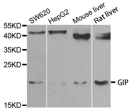
Anti-GIP Antibody
A31164
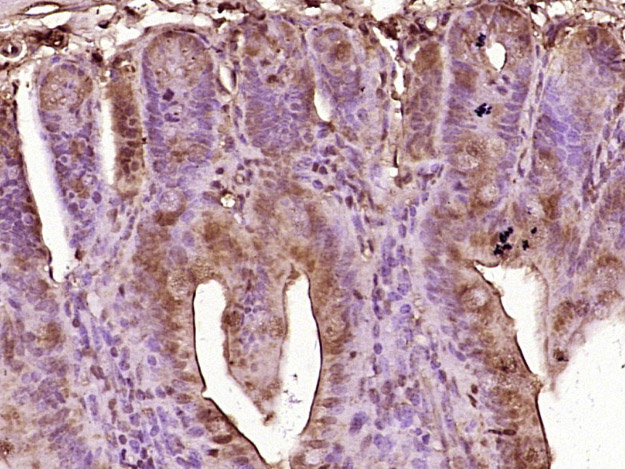
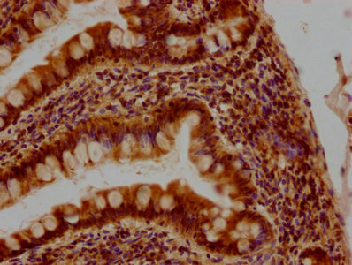
ApplicationsWestern Blot, ImmunoHistoChemistry
Product group Antibodies
ReactivityHuman, Mouse, Rat
Overview
- SupplierAntibodies.com
- Product NameAnti-GIP Antibody
- Delivery Days Customer7
- ApplicationsWestern Blot, ImmunoHistoChemistry
- CertificationResearch Use Only
- ClonalityPolyclonal
- ConjugateUnconjugated
- Estimated Purity>95%
- HostRabbit
- Scientific DescriptionRabbit polyclonal antibody to GIP
- ReactivityHuman, Mouse, Rat
- UNSPSC12352203
Related products
Product group Antibodies
Anti-GIP Antibody130-10094
ApplicationsELISA
ReactivityHuman
TargetGIP
- SizePrice
Product group Antibodies
GIP Polyclonal AntibodyBS-0098R
ApplicationsImmunoFluorescence, Western Blot, ELISA, ImmunoCytoChemistry, ImmunoHistoChemistry, ImmunoHistoChemistry Frozen, ImmunoHistoChemistry Paraffin
ReactivityBovine, Human, Mouse, Porcine, Rat
TargetGIP
- SizePrice
Product group Antibodies
GIP AntibodyCSB-PA009434PA01HU
ApplicationsELISA, ImmunoHistoChemistry
ReactivityHuman
TargetGIP
- SizePrice
Product group Antibodies
GIP Polyclonal AntibodyCAC12877
ApplicationsWestern Blot, ELISA
ReactivityDrosophila
- SizePrice
Product group Antibodies
GIP antibodyGTX04612
ApplicationsImmunoFluorescence, Western Blot, ImmunoCytoChemistry, ImmunoHistoChemistry, ImmunoHistoChemistry Paraffin
ReactivityHuman, Rat
TargetGIP
- SizePrice
Product group Antibodies

ApplicationsELISA
ReactivityHuman
TargetGIP
- SizePrice
Product group Antibodies
Anti-GIP AntibodyHPA021612
ApplicationsImmunoHistoChemistry
ReactivityHuman
TargetGIP
- SizePrice
Product group Antibodies
Anti-GIP AntibodyCAB6230
ApplicationsWestern Blot, ELISA, ImmunoHistoChemistry, ImmunoHistoChemistry Paraffin
ReactivityHuman
TargetGIP
- SizePrice

Product group Antibodies
Anti-GIP Antibody Picoband(r)PB9946-CARRIER-FREE
ApplicationsWestern Blot, ImmunoHistoChemistry
ReactivityHuman, Rat
TargetGIP
- SizePrice