


Immunohistochemical staining of human heart muscle shows moderate membranous and cytoplasmic positivity in myocytes.
Immunohistochemical staining of human heart muscle shows moderate membranous and cytoplasmic positivity in myocytes.
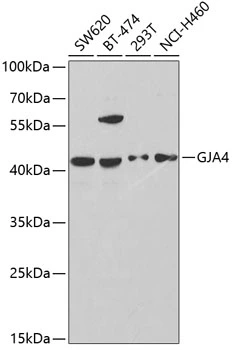
Anti-GJA4 Antibody
HPA047981
ApplicationsImmunoHistoChemistry
Product group Antibodies
ReactivityHuman
TargetGJA4
Overview
- SupplierAtlas Antibodies
- Product NameAnti-GJA4 Antibody
- Delivery Days Customer4
- ApplicationsImmunoHistoChemistry
- CertificationResearch Use Only
- ClonalityPolyclonal
- ConjugateUnconjugated
- Gene ID2701
- Target nameGJA4
- Target descriptiongap junction protein alpha 4
- Target synonymsCX37, gap junction alpha-4 protein, connexin-37, gap junction protein, alpha 4, 37kDa
- HostRabbit
- IsotypeIgG
- Protein IDP35212
- Protein NameGap junction alpha-4 protein
- Scientific DescriptionRecombinant Protein Epitope Signature Tag (PrEST) antigen sequence
- ReactivityHuman
- Storage Instruction-20°C,2°C to 8°C
- UNSPSC41116161
Datasheet
MSDS
Related products

Product group Antibodies
Anti-GJA4 AntibodyA98129
ApplicationsWestern Blot, ELISA
ReactivityHuman, Mouse, Rat
- SizePrice

Product group Antibodies
Anti-GJA4 Antibody Picoband(r)A05569-1-CARRIER-FREE
ApplicationsFlow Cytometry, Western Blot, ELISA
ReactivityHuman, Mouse, Rat
TargetGJA4
- SizePrice
Product group Antibodies
Anti-GJA4 Antibody144-02529
ApplicationsWestern Blot
ReactivityHuman
TargetGJA4
- SizePrice
Product group Antibodies
Connexin-37 Polyclonal AntibodyBS-4067R
ApplicationsImmunoFluorescence, Western Blot, ELISA, ImmunoCytoChemistry, ImmunoHistoChemistry, ImmunoHistoChemistry Frozen, ImmunoHistoChemistry Paraffin
ReactivityBovine, Equine, Guinea Pig, Human, Mouse, Porcine, Rabbit, Rat
TargetGJA4
- SizePrice
Product group Antibodies
GJA4 AntibodyCSB-PA008834
ApplicationsWestern Blot, ELISA
ReactivityHuman, Mouse, Rat
TargetGJA4
- SizePrice
Product group Antibodies
ApplicationsImmunoPrecipitation, Western Blot, ImmunoCytoChemistry, ImmunoHistoChemistry
ReactivityMouse, Rat
TargetGJA4
- SizePrice
Product group Antibodies

GJA4 / CX37 / Connexin 37 AntibodyLS-C332143
ApplicationsWestern Blot
ReactivityHuman
TargetGJA4
- SizePrice
Product group Antibodies
GJA4 antibodyGTX54119
ApplicationsWestern Blot
ReactivityHuman
TargetGJA4
- SizePrice