


Immunofluorescence staining of mouse periaqueductal grey shows immunoreactivity in neuronal cell bodies.
Immunofluorescence staining of mouse periaqueductal grey shows immunoreactivity in neuronal cell bodies.
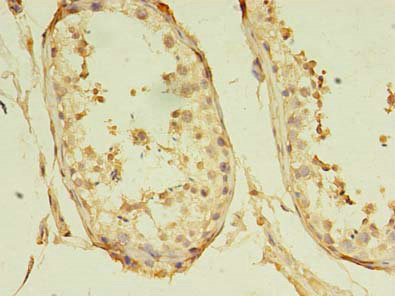
Anti-GKAP1 Antibody
HPA035117
ApplicationsImmunoHistoChemistry
Product group Antibodies
ReactivityHuman, Mouse
TargetGKAP1
Overview
- SupplierAtlas Antibodies
- Product NameAnti-GKAP1 Antibody
- Delivery Days Customer4
- ApplicationsImmunoHistoChemistry
- CertificationResearch Use Only
- ClonalityPolyclonal
- ConjugateUnconjugated
- Gene ID80318
- Target nameGKAP1
- Target descriptionG kinase anchoring protein 1
- Target synonymsFKSG21, GKAP42, G kinase-anchoring protein 1, cGMP-dependent protein kinase anchoring protein 42kDa, cGMP-dependent protein kinase-anchoring protein of 42 kDa, protein kinase anchoring protein GKAP42
- HostRabbit
- IsotypeIgG
- Protein IDQ5VSY0
- Protein NameG kinase-anchoring protein 1
- Scientific DescriptionRecombinant Protein Epitope Signature Tag (PrEST) antigen sequence
- ReactivityHuman, Mouse
- Storage Instruction-20°C,2°C to 8°C
- UNSPSC41116161
Datasheet
MSDS
Related products

Product group Antibodies
Anti-GKAP1 Antibody Picoband(r)A14397-1-CARRIER-FREE
ApplicationsFlow Cytometry, Western Blot, ELISA
ReactivityHuman, Mouse, Rat
TargetGKAP1
- SizePrice
Product group Antibodies

GKAP1 AntibodyLS-C830600
ApplicationsELISA, ImmunoHistoChemistry
ReactivityHuman, Mouse, Rat
TargetGKAP1
- SizePrice

Product group Antibodies
Anti-GKAP1 AntibodyHPA035118
ApplicationsWestern Blot, ImmunoCytoChemistry, ImmunoHistoChemistry
ReactivityHuman
TargetGKAP1
- SizePrice

Product group Antibodies
Anti-GKAP1 AntibodyHPA066173
ApplicationsWestern Blot, ImmunoHistoChemistry
ReactivityHuman
TargetGKAP1
- SizePrice
Product group Antibodies
GKAP1 AntibodyCSB-PA719617LA01HU
ApplicationsELISA, ImmunoHistoChemistry
ReactivityHuman
TargetGKAP1
- SizePrice
Product group Antibodies
GKAP1 AntibodyPACO36418
ApplicationsELISA, ImmunoHistoChemistry
ReactivityHuman
TargetGKAP1
- SizePrice
Product group Antibodies
GKAP1 Polyclonal AntibodyBS-13367R
ApplicationsImmunoFluorescence, Western Blot, ELISA, ImmunoCytoChemistry, ImmunoHistoChemistry, ImmunoHistoChemistry Frozen, ImmunoHistoChemistry Paraffin
ReactivityCanine, Equine, Human, Mouse, Rat
- SizePrice