

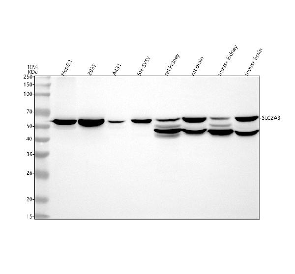
Figure 1. Western blot analysis of GLUT3 using anti-GLUT3 antibody (M03259). Electrophoresis was performed on a 5-20% SDS-PAGE gel at 70V (Stacking gel) / 90V (Resolving gel) for 2-3 hours. The sample well of each lane was loaded with 30 ug of sample under reducing conditions. Lane 1: human HepG2 whole cell lysates, Lane 2: human 293T whole cell lysates, Lane 3: human A431 whole cell lysates, Lane 4: human SH-SY5Y whole cell lysates, Lane 5: rat kidney tissue lysates, Lane 6: rat brain tissue lysates, Lane 7: mouse kidney tissue lysates, Lane 8: mouse brain tissue lysates. After electrophoresis, proteins were transferred to a nitrocellulose membrane at 150 mA for 50-90 minutes. Blocked the membrane with 5% non-fat milk/TBS for 1.5 hour at RT. The membrane was incubated with rabbit anti-GLUT3 antigen affinity purified monoclonal antibody (Catalog # M03259) at 1:500 overnight at 4°C, then washed with TBS-0.1%Tween 3 times with 5 minutes each and probed with a goat anti-rabbit IgG-HRP secondary antibody at a dilution of 1:500 for 1.5 hour at RT. The signal is developed using an Enhanced Chemiluminescent detection (ECL) kit (Catalog # EK1002) with Tanon 5200 system. A specific band was detected for GLUT3 at approximately 54 kDa. The expected band size for GLUT3 is at 54 kDa.
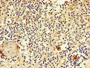
. GLUT3/SLC2A3 was detected in a paraffin-embedded section of human colon cancer tissue. Heat mediated antigen retrieval was performed in EDTA buffer (pH 8.0, epitope retrieval solution). The tissue section was blocked with 10% goat serum. The tissue section was then incubated with 1:200 rabbit anti-GLUT3/SLC2A3 Antibody (M03259) overnight at 4°C. Peroxidase Conjugated Goat Anti-rabbit IgG was used as secondary antibody and incubated for 30 minutes at 37°C. The tissue section was developed using HRP Conjugated Rabbit IgG Super Vision Assay Kit (Catalog # SV0002) with DAB as the chromogen.")
. GLUT3/SLC2A3 was detected in a paraffin-embedded section of human stomach cancer tissue. Heat mediated antigen retrieval was performed in EDTA buffer (pH 8.0, epitope retrieval solution). The tissue section was blocked with 10% goat serum. The tissue section was then incubated with 1:200 rabbit anti-GLUT3/SLC2A3 Antibody (M03259) overnight at 4°C. Peroxidase Conjugated Goat Anti-rabbit IgG was used as secondary antibody and incubated for 30 minutes at 37°C. The tissue section was developed using HRP Conjugated Rabbit IgG Super Vision Assay Kit (Catalog # SV0002) with DAB as the chromogen.")
Figure 1. Western blot analysis of GLUT3 using anti-GLUT3 antibody (M03259). Electrophoresis was performed on a 5-20% SDS-PAGE gel at 70V (Stacking gel) / 90V (Resolving gel) for 2-3 hours. The sample well of each lane was loaded with 30 ug of sample under reducing conditions. Lane 1: human HepG2 whole cell lysates, Lane 2: human 293T whole cell lysates, Lane 3: human A431 whole cell lysates, Lane 4: human SH-SY5Y whole cell lysates, Lane 5: rat kidney tissue lysates, Lane 6: rat brain tissue lysates, Lane 7: mouse kidney tissue lysates, Lane 8: mouse brain tissue lysates. After electrophoresis, proteins were transferred to a nitrocellulose membrane at 150 mA for 50-90 minutes. Blocked the membrane with 5% non-fat milk/TBS for 1.5 hour at RT. The membrane was incubated with rabbit anti-GLUT3 antigen affinity purified monoclonal antibody (Catalog # M03259) at 1:500 overnight at 4°C, then washed with TBS-0.1%Tween 3 times with 5 minutes each and probed with a goat anti-rabbit IgG-HRP secondary antibody at a dilution of 1:500 for 1.5 hour at RT. The signal is developed using an Enhanced Chemiluminescent detection (ECL) kit (Catalog # EK1002) with Tanon 5200 system. A specific band was detected for GLUT3 at approximately 54 kDa. The expected band size for GLUT3 is at 54 kDa.
Anti-GLUT3 SLC2A3 Rabbit Monoclonal Antibody
M03259
ApplicationsWestern Blot, ImmunoHistoChemistry
Product group Antibodies
ReactivityHuman, Mouse, Rat
TargetSLC2A3
Overview
- SupplierBoster Bio
- Product NameAnti-GLUT3 SLC2A3 Rabbit Monoclonal Antibody
- Delivery Days Customer9
- ApplicationsWestern Blot, ImmunoHistoChemistry
- CertificationResearch Use Only
- ClonalityMonoclonal
- Clone IDABHB-19
- Gene ID6515
- Target nameSLC2A3
- Target descriptionsolute carrier family 2 member 3
- Target synonymsGLUT3, solute carrier family 2, facilitated glucose transporter member 3, GLUT-3, glucose transporter type 3, brain, solute carrier family 2 (facilitated glucose transporter), member 3
- HostRabbit
- IsotypeIgG
- Protein IDP11169
- Protein NameSolute carrier family 2, facilitated glucose transporter member 3
- Scientific DescriptionBoster Bio Anti-GLUT3 SLC2A3 Rabbit Monoclonal Antibody catalog # M03259. Tested in WB, IHC application. This antibody reacts with Human, Mouse, Rat.
- ReactivityHuman, Mouse, Rat
- Storage Instruction-20°C
- UNSPSC12352203
Datasheet
MSDS
Related products
Product group Antibodies
SLC2A3 AntibodyCSB-PA021553LA01HU
ApplicationsImmunoFluorescence, ELISA, ImmunoHistoChemistry
ReactivityHuman
TargetSLC2A3
- SizePrice
Product group Antibodies
Anti-SLC2A3 Antibody144-66552
ApplicationsWestern Blot, ImmunoHistoChemistry
ReactivityHuman, Mouse
TargetSLC2A3
- SizePrice
Product group Antibodies
Anti-GLUT3 AntibodyA99645
ApplicationsWestern Blot, ELISA, ImmunoHistoChemistry
ReactivityHuman
- SizePrice

Product group Antibodies
Anti-SLC2A3 AntibodyHPA006539
ApplicationsImmunoCytoChemistry, ImmunoHistoChemistry
ReactivityHuman
TargetSLC2A3
- SizePrice
Product group Antibodies

SLC2A3 / GLUT3 AntibodyLS-C405625
ApplicationsWestern Blot, ELISA
ReactivityHuman
TargetSLC2A3
- SizePrice
Product group Antibodies
GLUT3 antibodyGTX129175
ApplicationsImmunoFluorescence, Western Blot, ImmunoCytoChemistry, ImmunoHistoChemistry
ReactivityHuman, Mouse, Rat
TargetSLC2A3
- SizePrice
Product group Antibodies
GLUT3 Recombinant Antibody, Biotin ConjugatedBSM-61510R-BIOTIN
ApplicationsWestern Blot
ReactivityHuman, Mouse, Rat
TargetSLC2A3
- SizePrice
Product group Antibodies
Slc2A3 Polyclonal AntibodyCAC10935
ApplicationsImmunoFluorescence, ELISA, ImmunoHistoChemistry
TargetSLC2A3
- SizePrice