


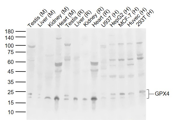
Figure 1. Western blot analysis of GPX4 using anti-GPX4 antibody (M02059). Electrophoresis was performed on a 5-20% SDS-PAGE gel at 70V (Stacking gel) / 90V (Resolving gel) for 2-3 hours. The sample well of each lane was loaded with 30 ug of sample under reducing conditions. Lane 1: human HepG2 whole cell lysates, Lane 2: human CACO-2 whole cell lysates, Lane 3: human 293T whole cell lysates, Lane 4: human K562 whole cell lysates, Lane 5: rat kidney tissue lysates, Lane 6: rat testis tissue lysates, Lane 7: mouse kidney tissue lysates, Lane 8: mouse testis tissue lysates. After electrophoresis, proteins were transferred to a nitrocellulose membrane at 150 mA for 50-90 minutes. Blocked the membrane with 5% non-fat milk/TBS for 1.5 hour at RT. The membrane was incubated with rabbit anti-GPX4 antigen affinity purified monoclonal antibody (Catalog # M02059) at 1:500 overnight at 4°C, then washed with TBS-0.1%Tween 3 times with 5 minutes each and probed with a goat anti-rabbit IgG-HRP secondary antibody at a dilution of 1:1000 for 1.5 hour at RT. The signal is developed using an Enhanced Chemiluminescent detection (ECL) kit (Catalog # EK1002) with Tanon 5200 system. A specific band was detected for GPX4 at approximately 19 kDa. The expected band size for GPX4 is at 22 kDa.

Figure 1. Western blot analysis of GPX4 using anti-GPX4 antibody (M02059). Electrophoresis was performed on a 5-20% SDS-PAGE gel at 70V (Stacking gel) / 90V (Resolving gel) for 2-3 hours. The sample well of each lane was loaded with 30 ug of sample under reducing conditions. Lane 1: human HepG2 whole cell lysates, Lane 2: human CACO-2 whole cell lysates, Lane 3: human 293T whole cell lysates, Lane 4: human K562 whole cell lysates, Lane 5: rat kidney tissue lysates, Lane 6: rat testis tissue lysates, Lane 7: mouse kidney tissue lysates, Lane 8: mouse testis tissue lysates. After electrophoresis, proteins were transferred to a nitrocellulose membrane at 150 mA for 50-90 minutes. Blocked the membrane with 5% non-fat milk/TBS for 1.5 hour at RT. The membrane was incubated with rabbit anti-GPX4 antigen affinity purified monoclonal antibody (Catalog # M02059) at 1:500 overnight at 4°C, then washed with TBS-0.1%Tween 3 times with 5 minutes each and probed with a goat anti-rabbit IgG-HRP secondary antibody at a dilution of 1:1000 for 1.5 hour at RT. The signal is developed using an Enhanced Chemiluminescent detection (ECL) kit (Catalog # EK1002) with Tanon 5200 system. A specific band was detected for GPX4 at approximately 19 kDa. The expected band size for GPX4 is at 22 kDa.
Anti-GPX4 Monoclonal Antibody
M02059
ApplicationsImmunoFluorescence, Western Blot, ImmunoCytoChemistry, ImmunoHistoChemistry
Product group Antibodies
ReactivityHuman, Mouse, Rat
TargetGPX4
Overview
- SupplierBoster Bio
- Product NameAnti-GPX4 Monoclonal Antibody
- Delivery Days Customer9
- ApplicationsImmunoFluorescence, Western Blot, ImmunoCytoChemistry, ImmunoHistoChemistry
- CertificationResearch Use Only
- ClonalityMonoclonal
- Clone IDACCO-7
- Gene ID2879
- Target nameGPX4
- Target descriptionglutathione peroxidase 4
- Target synonymsGPx-4, GSHPx-4, MCSP, PHGPx, SMDS, snGPx, snPHGPx, phospholipid hydroperoxide glutathione peroxidase GPX4, epididymis secretory sperm binding protein, phospholipid hydroperoxidase, phospholipid hydroperoxide glutathione peroxidase, mitochondrial, selenoprotein GPX4, sperm nucleus glutathione peroxidase
- HostRabbit
- IsotypeIgG
- Protein IDP36969
- Protein NamePhospholipid hydroperoxide glutathione peroxidase GPX4
- Scientific DescriptionBoster Bio Anti-GPX4 Rabbit Monoclonal Antibody catalog # M02059. Tested in WB, IHC, ICC/IF applications. This antibody reacts with Human, Mouse, Rat.
- ReactivityHuman, Mouse, Rat
- Storage Instruction-20°C
- UNSPSC12352203
References
- Liu C, Wu C, Zhang S, et al. Contribution of impaired autophagy, mitochondrial dysfunction and abnormal lipolysis to epididymal aging in mice. Exp Gerontol. 2024,195:112528. doi: 10.1016/j.exger.2024.112528Read this paper
- Dai S, Kong H, Ja Y, et al. Expression of the laminin genes family and its relationship to prognosis in pancreatic carcinoma. Arab J Gastroenterol. 2024,25(3):306-314. doi: 10.1016/j.ajg.2024.07.006Read this paper
- Xiang X, Xu M, Liu L, et al. Liproxstatin-1 attenuates acute hypertriglyceridemic pancreatitis through inhibiting ferroptosis in rats. Sci Rep. 2024,14(1):9548. doi: 10.1038/s41598-024-60159-7Read this paper
- Liu X, Wang FY, Chi S, et al. Mitochondria-targeting peptide SS-31 attenuates ferroptosis via inhibition of the p38 MAPK signaling pathway in the hippocampus of epileptic rats. Brain Res. 2024,1836:148882. doi: 10.1016/j.brainres.2024.148882Read this paper
- Che H, Yi J, Zhao X, et al. Characterization of PKCα-rutin interactions and their application as a treatment strategy for pulmonary arterial hypertension by inhibiting ferroptosis. Food Funct. 2024,15(2):779-793. doi: 10.1039/d3fo01306eRead this paper
- Yi P, Huang Y, Zhao X, et al. A novel UVA-associated circUBE2I mediates ferroptosis in HaCaT cells. Photochem Photobiol. 2024,100(5):1365-1377. doi: 10.1111/php.13885Read this paper
- Liao J, Wei M, Wang J, et al. Naotaifang formula attenuates OGD/R-induced inflammation and ferroptosis by regulating microglial M1/M2 polarization through BMP6/SMADs signaling pathway. Biomed Pharmacother. 2023,167:115465. doi: 10.1016/j.biopha.2023.115465Read this paper
- Zhao F, Peng C, Li H, et al. Paeoniae Radix Rubra extract attenuates cerebral ischemia injury by inhibiting ferroptosis and activating autophagy through the PI3K/Akt signalling pathway. J Ethnopharmacol. 2023,315:116567. doi: 10.1016/j.jep.2023.116567Read this paper
- Li W, Fu H, Fang L, et al. Andrographolide induced ferroptosis in multiple myeloma cells by regulating the P38/Nrf2/HO-1 pathway. Arch Biochem Biophys. 2023,742:109622. doi: 10.1016/j.abb.2023.109622Read this paper
- Lv Y, Chen D, Tian X, et al. Protectin conjugates in tissue regeneration 1 alleviates sepsis-induced acute lung injury by inhibiting ferroptosis. J Transl Med. 2023,21(1):293. doi: 10.1186/s12967-023-04111-9Read this paper
Datasheet
MSDS
Related products
Product group Antibodies
Gpx4 Polyclonal AntibodyCAC07902
ApplicationsImmunoFluorescence, ELISA, ImmunoHistoChemistry
ReactivityMouse, Rat
TargetGPX4
- SizePrice
Product group Antibodies
GPX4 Polyclonal AntibodyBS-3884R
ApplicationsWestern Blot, ELISA
ReactivityBovine, Human, Mouse, Porcine, Rat, Sheep
TargetGPX4
- SizePrice
Product group Antibodies
Anti-Oct6 AntibodyA84999
ApplicationsWestern Blot, ELISA
ReactivityHuman
- SizePrice
Product group Antibodies
Anti-GPX4 Antibody144-01933
ApplicationsWestern Blot, ImmunoHistoChemistry
ReactivityHuman, Mouse, Rat
TargetGPX4
- SizePrice
Product group Antibodies
References

ApplicationsImmunoPrecipitation, Western Blot, ELISA, ImmunoHistoChemistry
ReactivityBovine, Human, Mouse, Porcine, Rat
TargetGPX4
- SizePrice
Product group Antibodies
GPX4 antibodyGTX04794
ApplicationsWestern Blot, ImmunoHistoChemistry, ImmunoHistoChemistry Paraffin
ReactivityHuman, Mouse, Rat
TargetGPX4
- SizePrice
Product group Antibodies

GPX4 / MCSP AntibodyLS-C748363
ApplicationsImmunoFluorescence, Western Blot, ImmunoHistoChemistry
ReactivityHuman, Mouse, Rat
TargetGPX4
- SizePrice