Bio-Connect
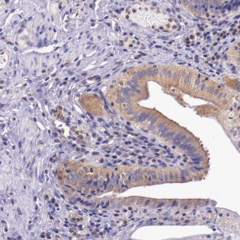
Immunohistochemical staining of human cerebral cortex shows moderate cytoplasmic positivity in neurons.

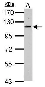
Anti-GRIPAP1 Antibody

HPA000615
Atlas Antibodies
ApplicationsWestern Blot, ImmunoCytoChemistry, ImmunoHistoChemistry
Product group Antibodies
TargetGRIPAP1
Sign in to order and to see your custom pricing.
Large volume orders?
Order with a bulk request

Overview

  • Supplier
    Atlas Antibodies
  • Product Name
    Anti-GRIPAP1
  • Delivery Days Customer
    4
  • Applications
    Western Blot, ImmunoCytoChemistry, ImmunoHistoChemistry
  • Certification
    Research Use Only
  • Clonality
    Polyclonal
  • Conjugate
    Unconjugated
  • Gene ID56850
  • Target name
    GRIPAP1
  • Target description
    GRIP1 associated protein 1
  • Target synonyms
    GRASP-1; GRIP1-associated protein 1
  • Host
    Rabbit
  • Isotype
    IgG
  • Protein IDQ4V328
  • Protein Name
    GRIP1-associated protein 1
  • Scientific Description
    Recombinant Protein Epitope Signature Tag (PrEST) antigen sequence
  • Storage Instruction
    -20°C,2°C to 8°C
  • UNSPSC
    12352203

Related products

Product group Antibodies
Everest Biotech
ApplicationsELISA, ImmunoHistoChemistry
TargetGRIPAP1
  • SizePrice
Figure 1. Western blot analysis of GRASP1/GRIPAP1 using anti-GRASP1/GRIPAP1 antibody (A13005-2). Electrophoresis was performed on a 5-20% SDS-PAGE gel at 70V (Stacking gel) / 90V (Resolving gel) for 2-3 hours. The sample well of each lane was loaded with 30 ug of sample under reducing conditions. Lane 1: human Hela whole cell lysates, Lane 2: huamn HepG2 whole cell lysates, Lane 3: human RT4 whole cell lysates, Lane 4: human MCF-7 whole cell lysates, Lane 5: human SiHa whole cell lysates, Lane 6: human T-47D whole cell lysates, Lane 7: rat brain tissue lysates, Lane 8: rat panceras tissue lysates, Lane 9: rat testis tissue lysates, Lane 10: rat PC-12 whole cell lysates, Lane 11: mouse brain tissue lysates, Lane 12: mouse pancreas tissue lysates, Lane 13: mouse NIH/3T3 whole cell lysates. After electrophoresis, proteins were transferred to a nitrocellulose membrane at 150 mA for 50-90 minutes. Blocked the membrane with 5% non-fat milk/TBS for 1.5 hour at RT. The membrane was incubated with rabbit anti-GRASP1/GRIPAP1 antigen affinity purified polyclonal antibody (Catalog # A13005-2) at 0.25 microg/mL overnight at 4°C, then washed with TBS-0.1%Tween 3 times with 5 minutes each and probed with a goat anti-rabbit IgG-HRP secondary antibody at a dilution of 1:5000 for 1.5 hour at RT. The signal is developed using an Enhanced Chemiluminescent detection (ECL) kit (Catalog # EK1002) with Tanon 5200 system. A specific band was detected for GRASP1/GRIPAP1 at approximately 96 kDa. The expected band size for GRASP1/GRIPAP1 is at 96 kDa.
Product group Antibodies
Boster Bio
ApplicationsFlow Cytometry, Western Blot, ELISA
TargetGRIPAP1
  • SizePrice
Product group Antibodies
GeneTex
ApplicationsWestern Blot
TargetGRIPAP1
  • SizePrice
Immunohistochemical staining of human gall bladder shows cytoplasmic positivity in glandular cells.
Product group Antibodies
Atlas Antibodies
ApplicationsImmunoHistoChemistry
TargetGRIPAP1
  • SizePrice
Product group Antibodies
Cusabio
GRIPAP1 AntibodyCSB-PA009923GA01HU
ApplicationsWestern Blot, ELISA, ImmunoHistoChemistry
TargetGRIPAP1
  • SizePrice
Product group Antibodies
Bethyl Laboratories
ApplicationsImmunoPrecipitation, Western Blot
TargetGRIPAP1
  • SizePrice
Product group Antibodies
RayBiotech
ApplicationsWestern Blot
TargetGRIPAP1
  • SizePrice
Atlas Antibodies
Protein IDQ4V328
  • SizePrice
Atlas Antibodies
Protein IDQ4V328
  • SizePrice
Product group Antibodies
LifeSpan BioSciences
ApplicationsELISA, ImmunoHistoChemistry
TargetGRIPAP1
  • SizePrice