


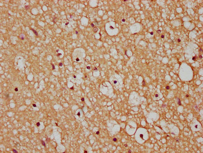
Immunohistochemistry analysis in human cerebral cortex and skeletal muscle tissues using Anti-HACD3 antibody. Corresponding HACD3 RNA-seq data are presented for the same tissues.
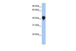
![Lane 1: Marker [kDa] 250, 130, 100, 70, 55, 35, 25, 15, 10. Lane 2: Human cell line RT-4](https://atlasantibodies.s3.amazonaws.com/images/wb/hpa014837-wb-1.jpg "Lane 1: Marker [kDa] 250, 130, 100, 70, 55, 35, 25, 15, 10. Lane 2: Human cell line RT-4")
Immunohistochemistry analysis in human cerebral cortex and skeletal muscle tissues using Anti-HACD3 antibody. Corresponding HACD3 RNA-seq data are presented for the same tissues.
Anti-HACD3 Antibody
HPA014837
ApplicationsWestern Blot, ImmunoCytoChemistry, ImmunoHistoChemistry
Product group Antibodies
TargetHACD3
Overview
- SupplierAtlas Antibodies
- Product NameAnti-HACD3
- Delivery Days Customer4
- ApplicationsWestern Blot, ImmunoCytoChemistry, ImmunoHistoChemistry
- CertificationResearch Use Only
- ClonalityPolyclonal
- ConjugateUnconjugated
- Gene ID51495
- Target nameHACD3
- Target description3-hydroxyacyl-CoA dehydratase 3
- Target synonymsBIND1; B-IND1; butyrate-induced protein 1; butyrate-induced transcript 1; HSPC121; protein tyrosine phosphatase-like A domain containing 1; protein tyrosine phosphatase-like protein PTPLAD1; protein-tyrosine phosphatase-like A domain-containing protein 1; PTPLAD1; very-long-chain (3R)-3-hydroxyacyl-[acyl-carrier protein] dehydratase 3; very-long-chain (3R)-3-hydroxyacyl-CoA dehydratase 3
- HostRabbit
- IsotypeIgG
- Protein IDQ9P035
- Protein NameVery-long-chain (3R)-3-hydroxyacyl-CoA dehydratase 3
- Scientific DescriptionRecombinant Protein Epitope Signature Tag (PrEST) antigen sequence
- Storage Instruction-20°C,2°C to 8°C
- UNSPSC12352203
Related products
Product group Antibodies
HACD3 AntibodyCSB-PA878945LA01HU
ApplicationsELISA, ImmunoHistoChemistry
TargetHACD3
- SizePrice

Product group Antibodies
Anti-HACD3 AntibodyHPA060347
ApplicationsImmunoCytoChemistry
TargetHACD3
- SizePrice
Product group Antibodies
Anti-HACD3 AntibodyHPA060347
ApplicationsImmunoCytoChemistry
TargetHACD3
- SizePrice
Product group Antibodies
PTPLAD1 antibody, N-termGTX46341
ApplicationsWestern Blot
TargetHACD3
- SizePrice
Product group Antibodies

HACD3 / PTPLAD1 Antibody (aa32-58)LS-C158767
ApplicationsWestern Blot
TargetHACD3
- SizePrice
Product group Antibodies
Anti-PTPLAD1 (N-term) Antibody102-23379
ApplicationsWestern Blot
TargetHACD3
- SizePrice