


Immunofluorescent staining of human cell line MCF7 shows localization to mitochondria.

Immunofluorescent staining of human cell line MCF7 shows localization to mitochondria.
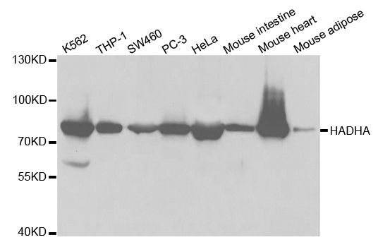
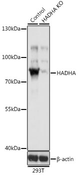
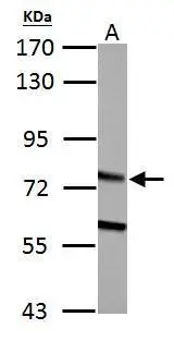
Anti-HADHA Antibody
HPA056070
ApplicationsWestern Blot, ImmunoCytoChemistry
Product group Antibodies
ReactivityHuman
TargetHADHA
Overview
- SupplierAtlas Antibodies
- Product NameAnti-HADHA Antibody
- Delivery Days Customer4
- ApplicationsWestern Blot, ImmunoCytoChemistry
- CertificationResearch Use Only
- ClonalityPolyclonal
- ConjugateUnconjugated
- Gene ID3030
- Target nameHADHA
- Target descriptionhydroxyacyl-CoA dehydrogenase trifunctional multienzyme complex subunit alpha
- Target synonymsECHA, GBP, HADH, LCEH, LCHAD, MLCL AT, MTPA, TP-ALPHA, trifunctional enzyme subunit alpha, mitochondrial, 3-ketoacyl-Coenzyme A (CoA) thiolase, alpha subunit, 3-oxoacyl-CoA thiolase, 78 kDa gastrin-binding protein, gastrin-binding protein, hydroxyacyl-CoA dehydrogenase/3-ketoacyl-CoA thiolase/enoyl-CoA hydratase (trifunctional protein), alpha subunit, hydroxyacyl-Coenzyme A dehydrogenase/3-ketoacyl-Coenzyme A thiolase/enoyl-Coenzyme A hydratase (trifunctional protein), alpha subunit, long-chain 2-enoyl-CoA hydratase, long-chain-3-hydroxyacyl-CoA dehydrogenase, mitochondrial long-chain 2-enoyl-Coenzyme A (CoA) hydratase, alpha subunit, mitochondrial long-chain L-3-hydroxyacyl-Coenzyme A (CoA) dehydrogenase, alpha subunit, mitochondrial trifunctional enzyme, alpha subunit, mitochondrial trifunctional protein, alpha subunit, monolysocardiolipin acyltransferase
- HostRabbit
- IsotypeIgG
- Protein IDP40939
- Protein NameTrifunctional enzyme subunit alpha, mitochondrial
- Scientific DescriptionRecombinant Protein Epitope Signature Tag (PrEST) antigen sequence
- ReactivityHuman
- Storage Instruction-20°C,2°C to 8°C
- UNSPSC41116161
Datasheet
MSDS
Related products
Product group Antibodies
Anti-HADHA AntibodyA30745
ApplicationsImmunoFluorescence, Western Blot, ImmunoHistoChemistry
ReactivityHuman, Mouse, Rat
- SizePrice

Product group Antibodies
Anti-HADHA Antibody Picoband(r)A03666-2-CARRIER-FREE
ApplicationsFlow Cytometry, ImmunoFluorescence, Western Blot, ELISA, ImmunoCytoChemistry, ImmunoHistoChemistry
ReactivityHuman, Mouse, Rat
TargetHADHA
- SizePrice
Product group Antibodies
Anti-HADHA Antibody144-05346
ApplicationsImmunoFluorescence, Western Blot
ReactivityHuman, Mouse
TargetHADHA
- SizePrice
Product group Antibodies
HADHA Polyclonal AntibodyBS-55092R
ApplicationsImmunoFluorescence, ImmunoPrecipitation, Western Blot, ImmunoCytoChemistry
ReactivityHuman, Mouse, Rat
TargetHADHA
- SizePrice
Product group Antibodies
HADHA AntibodyCSB-PA010118GA01HU
ApplicationsWestern Blot, ELISA, ImmunoHistoChemistry
ReactivityHuman, Mouse, Rat
TargetHADHA
- SizePrice
Product group Antibodies
HADHA antibodyGTX101177
ApplicationsImmunoFluorescence, Western Blot, ImmunoCytoChemistry, ImmunoHistoChemistry, ImmunoHistoChemistry Paraffin
ReactivityHuman, Mouse, Rat
TargetHADHA
- SizePrice
Product group Antibodies

HADHA AntibodyLS-C482426
ApplicationsImmunoFluorescence, Western Blot, ImmunoCytoChemistry, ImmunoHistoChemistry, ImmunoHistoChemistry Paraffin
ReactivityHuman, Mouse, Rat
TargetHADHA
- SizePrice