


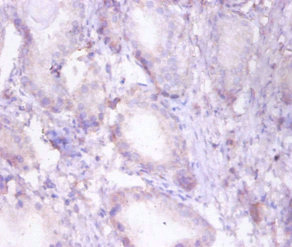
Immunohistochemical staining of human heart muscle shows strong granular cytoplasmic positivity in cardiomyocytes.
 shows similar pattern to independent antibody HPA037540 (B).")
Immunohistochemical staining of human heart muscle shows strong granular cytoplasmic positivity in cardiomyocytes.
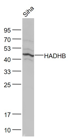
Anti-HADHB Antibody
HPA037539
ApplicationsWestern Blot, ImmunoHistoChemistry
Product group Antibodies
ReactivityHuman, Mouse, Rat
TargetHADHB
Overview
- SupplierAtlas Antibodies
- Product NameAnti-HADHB Antibody
- Delivery Days Customer4
- ApplicationsWestern Blot, ImmunoHistoChemistry
- CertificationResearch Use Only
- ClonalityPolyclonal
- ConjugateUnconjugated
- Gene ID3032
- Target nameHADHB
- Target descriptionhydroxyacyl-CoA dehydrogenase trifunctional multienzyme complex subunit beta
- Target synonymsECHB, MSTP029, MTPB, MTPD, MTPD2, TP-BETA, trifunctional enzyme subunit beta, mitochondrial, 2-enoyl-Coenzyme A (CoA) hydratase, beta subunit, 3-ketoacyl-Coenzyme A (CoA) thiolase of mitochondrial trifunctional protein, beta subunit, acetyl-CoA acyltransferase, beta-ketothiolase, hydroxyacyl-CoA dehydrogenase/3-ketoacyl-CoA thiolase/enoyl-CoA hydratase (trifunctional protein), beta subunit, hydroxyacyl-Coenzyme A dehydrogenase/3-ketoacyl-Coenzyme A thiolase/enoyl-Coenzyme A hydratase (trifunctional protein), beta subunit
- HostRabbit
- IsotypeIgG
- Protein IDP55084
- Protein NameTrifunctional enzyme subunit beta, mitochondrial
- Scientific DescriptionRecombinant Protein Epitope Signature Tag (PrEST) antigen sequence
- ReactivityHuman, Mouse, Rat
- Storage Instruction-20°C,2°C to 8°C
- UNSPSC41116161
Datasheet
MSDS
Related products

Product group Antibodies
Anti-HADHB Antibody Picoband(r)A04776-2-CARRIER-FREE
ApplicationsFlow Cytometry, ImmunoFluorescence, Western Blot, ELISA, ImmunoCytoChemistry, ImmunoHistoChemistry
ReactivityHuman, Mouse, Rat
TargetHADHB
- SizePrice
Product group Antibodies
Anti-HADHB AntibodyA31652
ApplicationsImmunoFluorescence, Western Blot, ImmunoHistoChemistry
ReactivityHuman, Mouse, Rat
- SizePrice
Product group Antibodies
Anti-HADHB Antibody144-05716
ApplicationsImmunoFluorescence, Western Blot
ReactivityHuman, Mouse, Rat
TargetHADHB
- SizePrice
Product group Antibodies
HADHB Polyclonal AntibodyBS-5065R
ApplicationsImmunoFluorescence, Western Blot, ELISA, ImmunoCytoChemistry, ImmunoHistoChemistry, ImmunoHistoChemistry Frozen, ImmunoHistoChemistry Paraffin
ReactivityBovine, Canine, Human, Mouse, Porcine, Rabbit, Rat, Sheep
TargetHADHB
- SizePrice
Product group Antibodies
HADHB Polyclonal AntibodyCAC13256
ApplicationsELISA, ImmunoHistoChemistry
TargetHADHB
- SizePrice
Product group Antibodies
HADHB AntibodyCSB-PA03035A0RB
ApplicationsELISA, ImmunoHistoChemistry
ReactivityHuman
TargetHADHB
- SizePrice
![Various whole cell extracts (30 μg) were separated by 10% SDS-PAGE, and the membrane was blotted with HADHB antibody [N1], N-term (GTX100302) diluted at 1:1000. The HRP-conjugated anti-rabbit IgG antibody (GTX213110-01) was used to detect the primary antibody.](https://www.genetex.com/upload/website/prouct_img/normal/GTX100302/GTX100302_39443_20220121_WB_w_23060100_143.webp)
Product group Antibodies
HADHB antibody [N1], N-termGTX100302
ApplicationsWestern Blot
ReactivityHuman
TargetHADHB
- SizePrice