

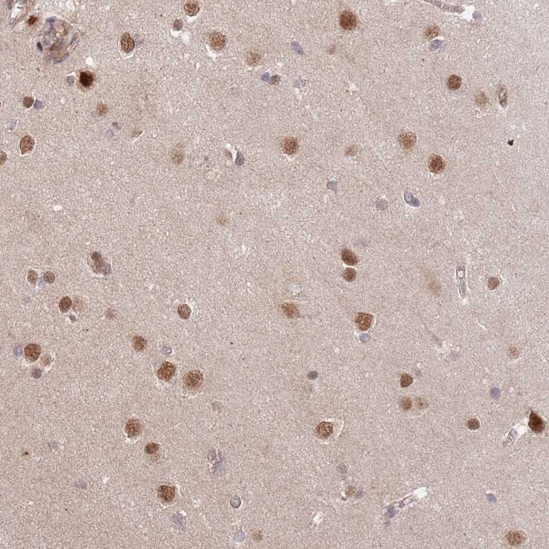
Immunohistochemical staining of human cerebral cortex shows moderate nuclear positivity in neuronal cells.
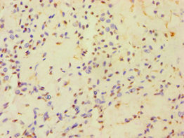
Immunohistochemical staining of human cerebral cortex shows moderate nuclear positivity in neuronal cells.
Anti-HAUS3 Antibody
HPA048190
ApplicationsImmunoHistoChemistry
Product group Antibodies
ReactivityHuman
TargetHAUS3
Overview
- SupplierAtlas Antibodies
- Product NameAnti-HAUS3 Antibody
- Delivery Days Customer4
- ApplicationsImmunoHistoChemistry
- CertificationResearch Use Only
- ClonalityPolyclonal
- ConjugateUnconjugated
- Gene ID79441
- Target nameHAUS3
- Target descriptionHAUS augmin like complex subunit 3
- Target synonymsC4orf15, IT1, dgt3, HAUS augmin-like complex subunit 3
- HostRabbit
- IsotypeIgG
- Protein IDQ68CZ6
- Protein NameHAUS augmin-like complex subunit 3
- Scientific DescriptionRecombinant Protein Epitope Signature Tag (PrEST) antigen sequence
- ReactivityHuman
- Storage Instruction-20°C,2°C to 8°C
- UNSPSC41116161
Datasheet
MSDS
Related products

Product group Antibodies
Anti-HAUS3 Antibody Picoband(r)A12796-2-CARRIER-FREE
ApplicationsFlow Cytometry, ImmunoFluorescence, Western Blot, ELISA, ImmunoCytoChemistry
ReactivityHuman, Mouse, Rat
TargetHAUS3
- SizePrice
Product group Antibodies
HAUS3 Polyclonal AntibodyBS-15414R
ApplicationsFlow Cytometry, ImmunoFluorescence, ELISA, ImmunoCytoChemistry, ImmunoHistoChemistry, ImmunoHistoChemistry Frozen, ImmunoHistoChemistry Paraffin
ReactivityHuman, Mouse, Rat
- SizePrice
Product group Antibodies
HAUS3 AntibodyCSB-PA734652ESR2HU
ApplicationsELISA, ImmunoHistoChemistry
ReactivityHuman
TargetHAUS3
- SizePrice
Product group Antibodies

HAUS3 / C4orf15 AntibodyLS-C409794
ApplicationsWestern Blot
ReactivityHuman, Mouse, Rat
TargetHAUS3
- SizePrice
Product group Antibodies
Anti-HAUS3 AntibodyHPA040649
ApplicationsImmunoCytoChemistry
ReactivityHuman
TargetHAUS3
- SizePrice