


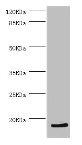
Figure 1. Western blot analysis of HBZ using anti-HBZ antibody (A03815-1). Electrophoresis was performed on a 5-20% SDS-PAGE gel at 70V (Stacking gel) / 90V (Resolving gel) for 2-3 hours. The sample well of each lane was loaded with 30 ug of sample under reducing conditions. Lane 1: rat bone tissue lysates, Lane 2: mouse bone tissue lysates. After electrophoresis, proteins were transferred to a nitrocellulose membrane at 150 mA for 50-90 minutes. Blocked the membrane with 5% non-fat milk/TBS for 1.5 hour at RT. The membrane was incubated with rabbit anti-HBZ antigen affinity purified polyclonal antibody (A03815-1) at 0.5 microg/mL overnight at 4°C, then washed with TBS-0.1%Tween 3 times with 5 minutes each and probed with a goat anti-rabbit IgG-HRP secondary antibody at a dilution of 1:5000 for 1.5 hour at RT. The signal is developed using an Enhanced Chemiluminescent detection (ECL) kit (Catalog # EK1002) with Tanon 5200 system. A specific band was detected for HBZ at approximately 14 kDa. The expected band size for HBZ is at 16 kDa.
Figure 1. Western blot analysis of HBZ using anti-HBZ antibody (A03815-1). Electrophoresis was performed on a 5-20% SDS-PAGE gel at 70V (Stacking gel) / 90V (Resolving gel) for 2-3 hours. The sample well of each lane was loaded with 30 ug of sample under reducing conditions. Lane 1: rat bone tissue lysates, Lane 2: mouse bone tissue lysates. After electrophoresis, proteins were transferred to a nitrocellulose membrane at 150 mA for 50-90 minutes. Blocked the membrane with 5% non-fat milk/TBS for 1.5 hour at RT. The membrane was incubated with rabbit anti-HBZ antigen affinity purified polyclonal antibody (A03815-1) at 0.5 microg/mL overnight at 4°C, then washed with TBS-0.1%Tween 3 times with 5 minutes each and probed with a goat anti-rabbit IgG-HRP secondary antibody at a dilution of 1:5000 for 1.5 hour at RT. The signal is developed using an Enhanced Chemiluminescent detection (ECL) kit (Catalog # EK1002) with Tanon 5200 system. A specific band was detected for HBZ at approximately 14 kDa. The expected band size for HBZ is at 16 kDa.
Anti-HBZ Antibody Picoband(r)
A03815-1-FITC
ApplicationsWestern Blot, ELISA
Product group Antibodies
ReactivityMouse, Rat
TargetHBZ
Overview
- SupplierBoster Bio
- Product NameAnti-HBZ Antibody Picoband(r)
- Delivery Days Customer9
- ApplicationsWestern Blot, ELISA
- CertificationResearch Use Only
- ClonalityPolyclonal
- Concentration500 ug/ml
- ConjugateFITC
- Gene ID3050
- Target nameHBZ
- Target descriptionhemoglobin subunit zeta
- Target synonymsHBAZ, HBZ-T1, HBZ1, hemoglobin subunit zeta, hemoglobin zeta chain, hemoglobin, zeta, zeta-globin
- HostRabbit
- IsotypeIgG
- Protein IDP02008
- Protein NameHemoglobin subunit zeta
- Scientific DescriptionBoster Bio Anti-HBZ Antibody Picoband® catalog # A03815-1. Tested in WB, ELISA applications. This antibody reacts with Mouse, Rat. The brand Picoband indicates this is a premium antibody that guarantees superior quality, high affinity, and strong signals with minimal background in Western blot applications. Only our best-performing antibodies are designated as Picoband, ensuring unmatched performance.
- ReactivityMouse, Rat
- Storage Instruction-20°C,2°C to 8°C
- UNSPSC12352203
Related products
Product group Antibodies
HBZ Polyclonal AntibodyBS-22473R
ApplicationsWestern Blot
ReactivityHuman
TargetHBZ
- SizePrice
Product group Antibodies
HBZ Polyclonal AntibodyCAC13290
ApplicationsWestern Blot, ELISA
ReactivityVirus
- SizePrice
Product group Antibodies
Hemoglobin zeta antibody [N1C3]GTX106483
ApplicationsWestern Blot
ReactivityHuman
TargetHBZ
- SizePrice
Product group Antibodies

HBZ AntibodyLS-C830876
ApplicationsWestern Blot, ELISA, ImmunoHistoChemistry
ReactivityHuman
TargetHBZ
- SizePrice

Product group Antibodies
Anti-HBZ AntibodyHPA063268
ApplicationsImmunoCytoChemistry
ReactivityHuman
TargetHBZ
- SizePrice
Product group Antibodies
HBZ AntibodyCSB-PA010162DSR1HU
ApplicationsWestern Blot, ELISA, ImmunoHistoChemistry
ReactivityHuman
TargetHBZ
- SizePrice
Product group Antibodies
Anti-HBZ Antibody Picoband(r)A03815-1-CARRIER-FREE
ApplicationsWestern Blot, ELISA
ReactivityMouse, Rat
TargetHBZ
- SizePrice