

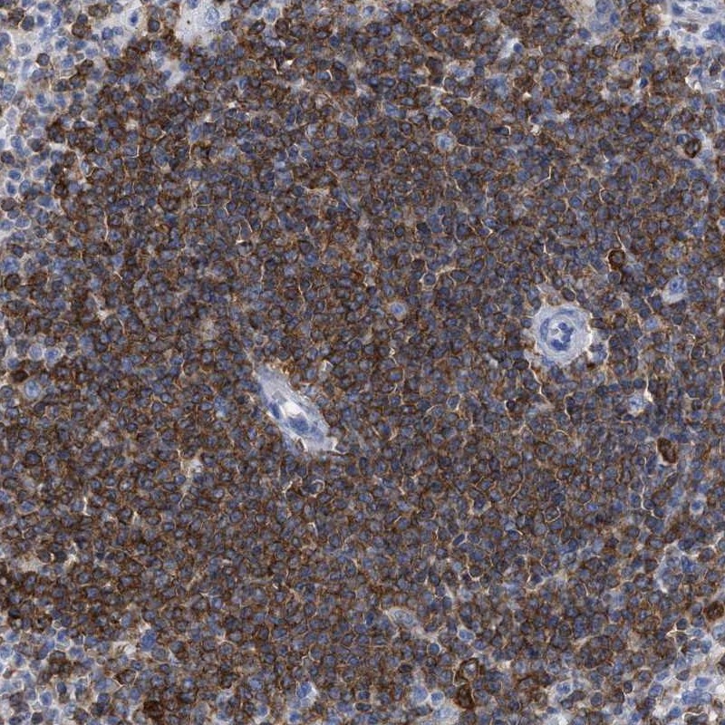
Immunohistochemical staining of human spleen shows strong cytoplasmic positivity in cells of white pulp.
Immunohistochemical staining of human spleen shows strong cytoplasmic positivity in cells of white pulp.
Anti-HCLS1 Antibody
HPA019143
ApplicationsImmunoHistoChemistry
Product group Antibodies
ReactivityHuman
TargetHCLS1
Overview
- SupplierAtlas Antibodies
- Product NameAnti-HCLS1 Antibody
- Delivery Days Customer4
- ApplicationsImmunoHistoChemistry
- CertificationResearch Use Only
- ClonalityPolyclonal
- ConjugateUnconjugated
- Gene ID3059
- Target nameHCLS1
- Target descriptionhematopoietic cell-specific Lyn substrate 1
- Target synonymsCTTNL, HS1, lckBP1, p75, hematopoietic lineage cell-specific protein
- HostRabbit
- IsotypeIgG
- Protein IDP14317
- Protein NameHematopoietic lineage cell-specific protein
- Scientific DescriptionRecombinant Protein Epitope Signature Tag (PrEST) antigen sequence
- ReactivityHuman
- Storage Instruction-20°C,2°C to 8°C
- UNSPSC41116161
Datasheet
MSDS
Related products
Product group Antibodies
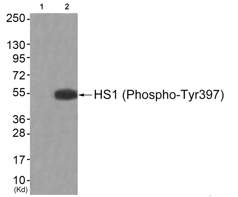
Phospho-HCLS1 (Y378) AntibodyCSB-PA009133
ApplicationsWestern Blot, ELISA
ReactivityHuman
TargetHCLS1
- SizePrice
Product group Antibodies
ApplicationsWestern Blot
ReactivityHuman
- SizePrice

Product group Antibodies
Anti-HCLS1 Antibody Picoband(r)A04313-1-CARRIER-FREE
ApplicationsFlow Cytometry, Western Blot, ELISA
ReactivityHuman
TargetHCLS1
- SizePrice
Product group Antibodies

HCLS1 AntibodyLS-C331948
ApplicationsWestern Blot, ImmunoHistoChemistry
ReactivityHuman, Mouse
TargetHCLS1
- SizePrice
Product group Antibodies
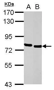
HCLS1 antibody [C3], C-termGTX100303
ApplicationsWestern Blot, ImmunoHistoChemistry, ImmunoHistoChemistry Paraffin
ReactivityHuman, Mouse
TargetHCLS1
- SizePrice
Product group Antibodies
Anti-HCLS1Y058085
ApplicationsWestern Blot, ELISA, ImmunoHistoChemistry
ReactivityHuman, Mouse
- SizePrice
Product group Antibodies
Anti-HCLS1 Antibody144-02165
ApplicationsWestern Blot, ImmunoHistoChemistry
ReactivityHuman, Mouse
TargetHCLS1
- SizePrice
Product group Antibodies
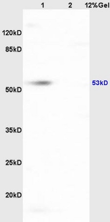
HCLS1 Polyclonal AntibodyBS-5109R
ApplicationsImmunoFluorescence, Western Blot, ELISA, ImmunoCytoChemistry, ImmunoHistoChemistry, ImmunoHistoChemistry Frozen, ImmunoHistoChemistry Paraffin
ReactivityBovine, Canine, Equine, Human, Mouse, Rabbit, Rat
TargetHCLS1
- SizePrice