


Immunofluorescent staining of human cell line MCF7 shows localization to nucleoli fibrillar center.
Immunofluorescent staining of human cell line MCF7 shows localization to nucleoli fibrillar center.
Anti-HDAC10 Antibody
HPA056514
ApplicationsImmunoCytoChemistry
Product group Antibodies
ReactivityHuman
TargetHDAC10
Overview
- SupplierAtlas Antibodies
- Product NameAnti-HDAC10 Antibody
- Delivery Days Customer4
- ApplicationsImmunoCytoChemistry
- CertificationResearch Use Only
- ClonalityPolyclonal
- ConjugateUnconjugated
- Gene ID83933
- Target nameHDAC10
- Target descriptionhistone deacetylase 10
- Target synonymsHD10, polyamine deacetylase HDAC10
- HostRabbit
- IsotypeIgG
- Protein IDQ969S8
- Protein NamePolyamine deacetylase HDAC10
- Scientific DescriptionRecombinant Protein Epitope Signature Tag (PrEST) antigen sequence
- ReactivityHuman
- Storage Instruction-20°C,2°C to 8°C
- UNSPSC41116161
Datasheet
MSDS
Related products
Product group Antibodies
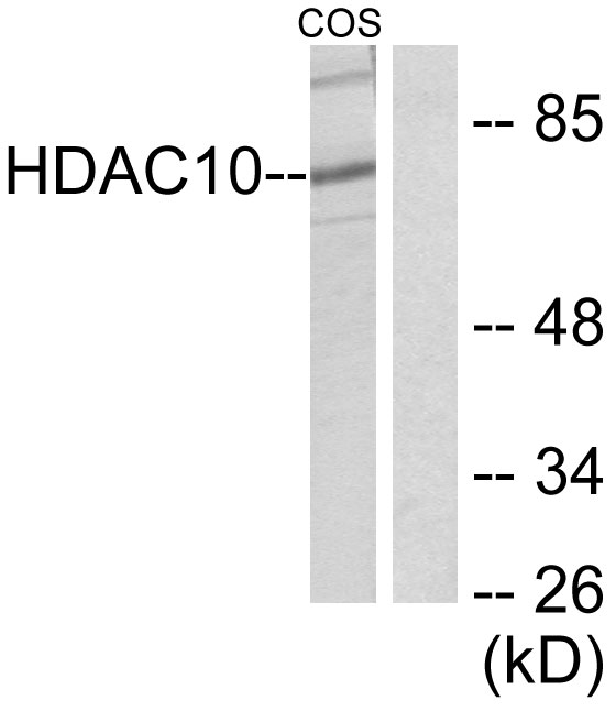
Anti-HDAC10 AntibodyA95624
ApplicationsWestern Blot, ELISA, ImmunoHistoChemistry
ReactivityHuman, Mouse, Rat
- SizePrice

Product group Antibodies
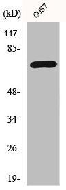
Anti-HDAC10 Antibody Picoband(r)A05215-2-CARRIER-FREE
ApplicationsWestern Blot, ELISA
ReactivityHuman, Mouse, Rat
TargetHDAC10
- SizePrice
Product group Antibodies
Anti-HDAC10 (N-term) Antibody102-20746
ApplicationsWestern Blot
TargetHDAC10
- SizePrice
Product group Antibodies
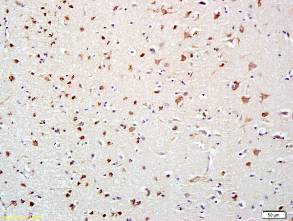
HDAC10 AntibodyCSB-PA002914
ApplicationsWestern Blot, ELISA, ImmunoHistoChemistry
ReactivityHuman, Monkey, Mouse, Rat
TargetHDAC10
- SizePrice
Product group Antibodies
HDAC10 Polyclonal AntibodyCAC14590
ApplicationsImmunoFluorescence, Western Blot, ELISA, ImmunoHistoChemistry
ReactivityMouse
TargetHDAC10
- SizePrice
Product group Antibodies
HDAC10 Polyclonal AntibodyBS-2893R
ApplicationsImmunoFluorescence, ELISA, ImmunoCytoChemistry, ImmunoHistoChemistry, ImmunoHistoChemistry Frozen, ImmunoHistoChemistry Paraffin
ReactivityBovine, Equine, Human, Mouse, Porcine, Rabbit, Rat
TargetHDAC10
- SizePrice
Product group Antibodies

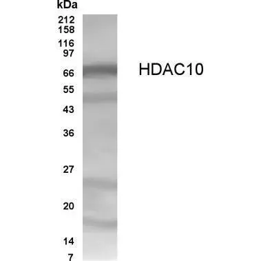
HDAC10 AntibodyLS-C408925
ApplicationsWestern Blot
ReactivityHuman, Mouse, Rat
TargetHDAC10
- SizePrice
Product group Antibodies
HDAC10 antibodyGTX70464
ApplicationsWestern Blot, ELISA
ReactivityHuman
TargetHDAC10
- SizePrice