

Anti-HDGF [H3]
Ab02457-1.1
ApplicationsImmunoFluorescence, ImmunoPrecipitation, Western Blot, ImmunoHistoChemistry, Neutralisation/Blocking
Product group Antibodies
ReactivityHuman, Mouse
TargetHDGF
Overview
- SupplierAbsolute Antibody
- Product NameAnti-HDGF [H3]
- Delivery Days Customer7
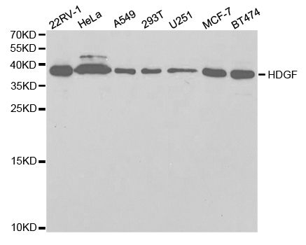
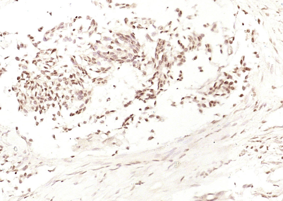
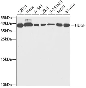
- Application Supplier NoteThis antibody was used for immunoprecipitation on total cell lysates from H460 NSCLC cells (US20070243191). The neutralizing ability of this antibody was tested on A549. The result was a significant decrease in tumor size (US20070243191). This antibody was fluorescently labelled and injected into nude mice to test the antibody distribution. The antibodies evenly distributed in the animals including the tumors. This antibody entered into the cytoplasmic compartment of some tumor cells (US20070243191). Immunohistochemistry with this antibody was performed on tissues from NSCLC patients (US20070243191). Western blot was performed on HDGF protein using this antibody (Ren et al, 2009;pmid:19435872). ELISA was performed on the HDGF protein using this antibody (US20070243191).
- ApplicationsImmunoFluorescence, ImmunoPrecipitation, Western Blot, ImmunoHistoChemistry, Neutralisation/Blocking
- Applications SupplierIP; IF; IHC; neutralizing; WB
- CertificationResearch Use Only
- ClonalityMonoclonal
- Clone IDH3
- Gene ID3068
- Target nameHDGF
- Target descriptionheparin binding growth factor
- Target synonymsHMG1L2, hepatoma-derived growth factor, epididymis secretory sperm binding protein, hepatoma derived growth factor, high mobility group protein 1-like 2
- HostMouse
- IsotypeIgG1
- Protein IDP51858
- Protein NameHepatoma-derived growth factor
- ReactivityHuman, Mouse
- Reactivity SupplierHuman, mouse
- Reactivity Supplier NoteThe original antibody was raised by immunizing mice with full-length recombinant HDGF. This antibody binds a different epitope on HDGF than antibody C1.
- Storage Instruction-20°C,2°C to 8°C
- UNSPSC41116161
Related products
Product group Antibodies
Anti-HDGF AntibodyA30746
ApplicationsImmunoFluorescence, ImmunoPrecipitation, Western Blot, ImmunoHistoChemistry, Other Application
ReactivityHuman, Mouse, Rat
- SizePrice

Product group Antibodies
Anti-HDGF Antibody Picoband(r)A01057-CARRIER-FREE
ApplicationsFlow Cytometry, ImmunoFluorescence, Western Blot, ImmunoCytoChemistry, ImmunoHistoChemistry
ReactivityHuman, Mouse, Rat
TargetHDGF
- SizePrice
Product group Antibodies
Anti-HDGF Antibody144-05347
ApplicationsWestern Blot, ImmunoHistoChemistry
ReactivityHuman, Mouse, Rat
TargetHDGF
- SizePrice
Product group Antibodies

HDGF AntibodyLS-C748691
ApplicationsImmunoFluorescence, Western Blot
ReactivityHuman
TargetHDGF
- SizePrice
Product group Antibodies
HDGF Polyclonal AntibodyBS-10956R
ApplicationsImmunoFluorescence, ELISA, ImmunoCytoChemistry, ImmunoHistoChemistry, ImmunoHistoChemistry Frozen, ImmunoHistoChemistry Paraffin
ReactivityBovine, Guinea Pig, Human, Mouse, Porcine, Rat
TargetHDGF
- SizePrice
Product group Antibodies
HDGF AntibodyCSB-PA01004A0RB
ApplicationsELISA
ReactivityHuman
TargetHDGF
- SizePrice
Product group Antibodies
ApplicationsImmunoPrecipitation, Western Blot, ImmunoCytoChemistry, ImmunoHistoChemistry
TargetHDGF
- SizePrice

Product group Antibodies
Anti-HDGF AntibodyHPA048728
ApplicationsImmunoCytoChemistry, ImmunoHistoChemistry
ReactivityHuman
TargetHDGF
- SizePrice
Product group Antibodies
HDGF antibodyGTX54144
ApplicationsImmunoFluorescence, Western Blot, ImmunoCytoChemistry, ImmunoHistoChemistry, ImmunoHistoChemistry Paraffin
ReactivityHuman, Mouse, Rat
TargetHDGF
- SizePrice
Product group Antibodies
ApplicationsImmunoFluorescence, Western Blot, ELISA, ImmunoCytoChemistry
ReactivityHuman
TargetHDGF
- SizePrice