

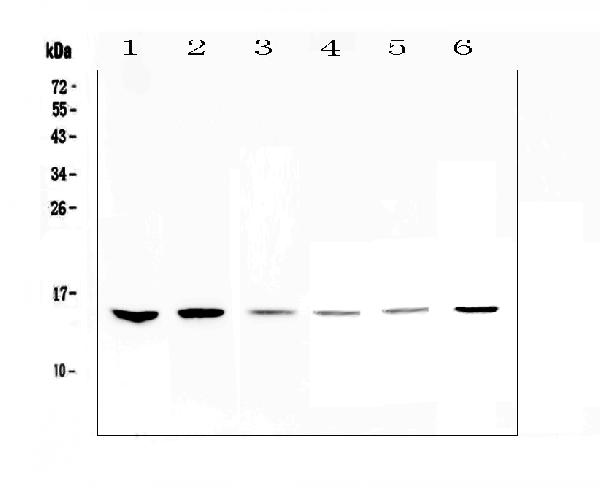
Figure 1. Western blot analysis of HE4 using anti-HE4 antibody (A02685-4). Electrophoresis was performed on a 5-20% SDS-PAGE gel at 70V (Stacking gel) / 90V (Resolving gel) for 2-3 hours. The sample well of each lane was loaded with 50ug of sample under reducing conditions. Lane 1: human Hela whole cell lysates, Lane 2: human MDA-MB-231 whole cell lysates, Lane 3: human MDA-MB-453 whole cell lysates, Lane 4: rat thymus tissue lysates, Lane 5: mouse testis tissue lysates, Lane 6: mouse thymus tissue lysates. After Electrophoresis, proteins were transferred to a Nitrocellulose membrane at 150mA for 50-90 minutes. Blocked the membrane with 5% Non-fat Milk/ TBS for 1.5 hour at RT. The membrane was incubated with rabbit anti-HE4 antigen affinity purified polyclonal antibody (Catalog # A02685-4) at 0.5 microg/mL overnight at 4°C, then washed with TBS-0.1%Tween 3 times with 5 minutes each and probed with a goat anti-rabbit IgG-HRP secondary antibody at a dilution of 1:10000 for 1.5 hour at RT. The signal is developed using an Enhanced Chemiluminescent detection (ECL) kit (Catalog # EK1002) with Tanon 5200 system. A specific band was detected for HE4 at approximately 15KD. The expected band size for HE4 is at 13KD.
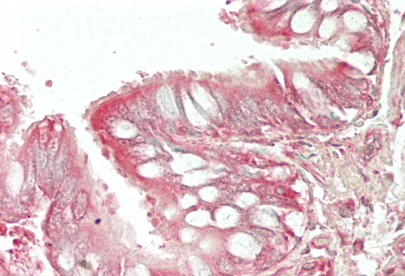
. HE4 was detected in paraffin-embedded section of rat small intestine tissue. Heat mediated antigen retrieval was performed in citrate buffer (pH6, epitope retrieval solution) for 20 mins. The tissue section was blocked with 10% goat serum. The tissue section was then incubated with 2microg/ml rabbit anti-HE4 Antibody (A02685-4) overnight at 4°C. Biotinylated goat anti-rabbit IgG was used as secondary antibody and incubated for 30 minutes at 37°C. The tissue section was developed using Strepavidin-Biotin-Complex (SABC)(Catalog # SA1022) with DAB as the chromogen.")
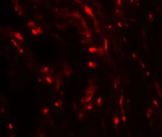
. HE4 was detected in paraffin-embedded section of mouse spleen tissue . Heat mediated antigen retrieval was performed in citrate buffer (pH6, epitope retrieval solution) for 20 mins. The tissue section was blocked with 10% goat serum. The tissue section was then incubated with 2microg/ml rabbit anti-HE4 Antibody (A02685-4) overnight at 4°C. Biotinylated goat anti-rabbit IgG was used as secondary antibody and incubated for 30 minutes at 37°C. The tissue section was developed using Strepavidin-Biotin-Complex (SABC)(Catalog # SA1022) with DAB as the chromogen.")
. HE4 was detected in paraffin-embedded section of rat spleen tissue . Heat mediated antigen retrieval was performed in citrate buffer (pH6, epitope retrieval solution) for 20 mins. The tissue section was blocked with 10% goat serum. The tissue section was then incubated with 2microg/ml rabbit anti-HE4 Antibody (A02685-4) overnight at 4°C. Biotinylated goat anti-rabbit IgG was used as secondary antibody and incubated for 30 minutes at 37°C. The tissue section was developed using Strepavidin-Biotin-Complex (SABC)(Catalog # SA1022) with DAB as the chromogen.")
Figure 1. Western blot analysis of HE4 using anti-HE4 antibody (A02685-4). Electrophoresis was performed on a 5-20% SDS-PAGE gel at 70V (Stacking gel) / 90V (Resolving gel) for 2-3 hours. The sample well of each lane was loaded with 50ug of sample under reducing conditions. Lane 1: human Hela whole cell lysates, Lane 2: human MDA-MB-231 whole cell lysates, Lane 3: human MDA-MB-453 whole cell lysates, Lane 4: rat thymus tissue lysates, Lane 5: mouse testis tissue lysates, Lane 6: mouse thymus tissue lysates. After Electrophoresis, proteins were transferred to a Nitrocellulose membrane at 150mA for 50-90 minutes. Blocked the membrane with 5% Non-fat Milk/ TBS for 1.5 hour at RT. The membrane was incubated with rabbit anti-HE4 antigen affinity purified polyclonal antibody (Catalog # A02685-4) at 0.5 microg/mL overnight at 4°C, then washed with TBS-0.1%Tween 3 times with 5 minutes each and probed with a goat anti-rabbit IgG-HRP secondary antibody at a dilution of 1:10000 for 1.5 hour at RT. The signal is developed using an Enhanced Chemiluminescent detection (ECL) kit (Catalog # EK1002) with Tanon 5200 system. A specific band was detected for HE4 at approximately 15KD. The expected band size for HE4 is at 13KD.
Anti-HE4/WFDC2 Antibody Picoband(r)
A02685-4-CARRIER-FREE
ApplicationsWestern Blot, ELISA, ImmunoHistoChemistry
Product group Antibodies
ReactivityHuman, Mouse, Rat
TargetWFDC2
Overview
- SupplierBoster Bio
- Product NameAnti-HE4/WFDC2 Antibody Picoband(r)
- Delivery Days Customer9
- ApplicationsWestern Blot, ELISA, ImmunoHistoChemistry
- CertificationResearch Use Only
- ClonalityPolyclonal
- Concentration500 ug/ml
- Gene ID10406
- Target nameWFDC2
- Target descriptionWAP four-disulfide core domain 2
- Target synonymsBENP, EDDM4, HE4, WAP5, dJ461P17.6, WAP four-disulfide core domain protein 2, WAP domain containing protein HE4-V4, epididymal protein 4, epididymal secretory protein E4, epididymis secretory sperm binding protein, epididymis-specific, whey-acidic protein type, four-disulfide core, human epididymis protein 4, major epididymis-specific protein E4, putative protease inhibitor WAP5
- HostRabbit
- IsotypeIgG
- Protein IDQ14508
- Protein NameWAP four-disulfide core domain protein 2
- Scientific DescriptionBoster Bio Anti-HE4/WFDC2 Antibody Picoband® catalog # A02685-4. Tested in ELISA, IHC, WB applications. This antibody reacts with Human, Mouse, Rat. The brand Picoband indicates this is a premium antibody that guarantees superior quality, high affinity, and strong signals with minimal background in Western blot applications. Only our best-performing antibodies are designated as Picoband, ensuring unmatched performance.
- ReactivityHuman, Mouse, Rat
- Storage Instruction-20°C,2°C to 8°C
- UNSPSC12352203
Related products
Product group Antibodies
Anti-HE4 AntibodyA286076
ApplicationsELISA, ImmunoHistoChemistry
ReactivityHuman, Mouse
- SizePrice
Product group Antibodies
ApplicationsWestern Blot, ELISA
ReactivityHuman
TargetWFDC2
- SizePrice

Product group Antibodies
Anti-WFDC2 AntibodyAMAB91819
ApplicationsImmunoHistoChemistry
ReactivityHuman
TargetWFDC2
- SizePrice
Product group Antibodies

ApplicationsWestern Blot, ELISA
ReactivityHuman
TargetWFDC2
- SizePrice
Product group Antibodies
HE4 Recombinant AntibodyBSM-52840R
ApplicationsFlow Cytometry, ImmunoFluorescence, Western Blot, ImmunoCytoChemistry, ImmunoHistoChemistry, ImmunoHistoChemistry Frozen, ImmunoHistoChemistry Paraffin
ReactivityHuman, Mouse
TargetWFDC2
- SizePrice
Product group Antibodies
WFDC2 AntibodyCSB-PA09794A0RB
ApplicationsImmunoFluorescence, Western Blot, ELISA, ImmunoHistoChemistry
ReactivityHuman, Mouse
TargetWFDC2
- SizePrice
Product group Antibodies

Goat anti-WFDC2 (aa29-43)EB09963
ApplicationsWestern Blot, ELISA
ReactivityCanine, Human
TargetWFDC2
- SizePrice
Product group Antibodies
Wfdc2 Polyclonal AntibodyCAC09115
ApplicationsImmunoFluorescence, Western Blot, ELISA, ImmunoHistoChemistry
ReactivityMouse
TargetWFDC2
- SizePrice
Product group Antibodies
HE4 antibodyGTX17207
ApplicationsWestern Blot, ELISA, ImmunoHistoChemistry, ImmunoHistoChemistry Paraffin
ReactivityHuman
TargetWFDC2
- SizePrice