


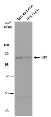
Figure 1. Western blot analysis of HIP1 using anti-HIP1 antibody (M02242). Electrophoresis was performed on a 5-20% SDS-PAGE gel at 70V (Stacking gel) / 90V (Resolving gel) for 2-3 hours. The sample well of each lane was loaded with 30 ug of sample under reducing conditions. Lane 1: human Hela whole cell lysates, Lane 2: human MCF-7 whole cell lysates, Lane 3: human PC-3 whole cell lysates, Lane 4: rat brain tissue lysates, Lane 5: rat PC-12 whole cell lysates, Lane 6: mouse brain tissue lysates, Lane 7: mouse NIH/3T3 whole cell lysates. After electrophoresis, proteins were transferred to a nitrocellulose membrane at 150 mA for 50-90 minutes. Blocked the membrane with 5% non-fat milk/TBS for 1.5 hour at RT. The membrane was incubated with rabbit anti-HIP1 antigen affinity purified monoclonal antibody (Catalog # M02242) at 1:1000 overnight at 4°C, then washed with TBS-0.1%Tween 3 times with 5 minutes each and probed with a goat anti-rabbit IgG-HRP secondary antibody at a dilution of 1:5000 for 1.5 hour at RT. The signal is developed using an Enhanced Chemiluminescent detection (ECL) kit (Catalog # EK1002) with Tanon 5200 system. A specific band was detected for HIP1 at approximately 116 kDa. The expected band size for HIP1 is at 116 kDa.
Figure 1. Western blot analysis of HIP1 using anti-HIP1 antibody (M02242). Electrophoresis was performed on a 5-20% SDS-PAGE gel at 70V (Stacking gel) / 90V (Resolving gel) for 2-3 hours. The sample well of each lane was loaded with 30 ug of sample under reducing conditions. Lane 1: human Hela whole cell lysates, Lane 2: human MCF-7 whole cell lysates, Lane 3: human PC-3 whole cell lysates, Lane 4: rat brain tissue lysates, Lane 5: rat PC-12 whole cell lysates, Lane 6: mouse brain tissue lysates, Lane 7: mouse NIH/3T3 whole cell lysates. After electrophoresis, proteins were transferred to a nitrocellulose membrane at 150 mA for 50-90 minutes. Blocked the membrane with 5% non-fat milk/TBS for 1.5 hour at RT. The membrane was incubated with rabbit anti-HIP1 antigen affinity purified monoclonal antibody (Catalog # M02242) at 1:1000 overnight at 4°C, then washed with TBS-0.1%Tween 3 times with 5 minutes each and probed with a goat anti-rabbit IgG-HRP secondary antibody at a dilution of 1:5000 for 1.5 hour at RT. The signal is developed using an Enhanced Chemiluminescent detection (ECL) kit (Catalog # EK1002) with Tanon 5200 system. A specific band was detected for HIP1 at approximately 116 kDa. The expected band size for HIP1 is at 116 kDa.
Anti-HIP1 Monoclonal Antibody
M02242
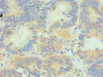
ApplicationsImmunoFluorescence, ImmunoPrecipitation, Western Blot, ImmunoCytoChemistry
Product group Antibodies
ReactivityHuman, Mouse, Rat
TargetHIP1
Overview
- SupplierBoster Bio
- Product NameAnti-HIP1 Monoclonal Antibody
- Delivery Days Customer9
- ApplicationsImmunoFluorescence, ImmunoPrecipitation, Western Blot, ImmunoCytoChemistry
- CertificationResearch Use Only
- ClonalityMonoclonal
- Clone IDAFG-8
- Gene ID3092
- Target nameHIP1
- Target descriptionhuntingtin interacting protein 1
- Target synonymsHIP-I, ILWEQ, SHON, SHONbeta, SHONgamma, huntingtin-interacting protein 1, huntingtin-interacting protein I
- HostRabbit
- IsotypeIgG
- Protein IDO00291
- Protein NameHuntingtin-interacting protein 1
- Scientific DescriptionBoster Bio Anti-HIP1 Monoclonal Antibody catalog # M02242. Tested in WB, ICC/IF, IP applications. This antibody reacts with Human, Mouse, Rat.
- ReactivityHuman, Mouse, Rat
- Storage Instruction-20°C
- UNSPSC12352203
References
- Zhang F, Xu X, Hou J, et al. Cardioprotective efficacy of Xin-shu-bao tablet in heart failure with reduced ejection fraction by modulating THBD/ARRB1/FGF1/STIM1 signaling. Biomed Pharmacother. 2023,165:115119. doi: 10.1016/j.biopha.2023.115119Read this paper
Datasheet
MSDS
Related products
Product group Antibodies
Anti-HIP1 AntibodyA31749
ApplicationsImmunoFluorescence, Western Blot, ImmunoHistoChemistry
ReactivityHuman, Mouse
- SizePrice
Product group Antibodies
Anti-HIP1 Antibody144-06921
ApplicationsImmunoFluorescence, Western Blot, ImmunoHistoChemistry
ReactivityHuman, Mouse
TargetHIP1
- SizePrice
Product group Antibodies
HIP1 Recombinant Antibody, AbBy Fluor-594 ConjugatedBSM-61799R-BF594
ApplicationsWestern Blot
ReactivityHuman, Mouse, Rat
TargetHIP1
- SizePrice
Product group Antibodies
HIP1 AntibodyCSB-PA010365ESR1HU
ApplicationsELISA, ImmunoHistoChemistry
ReactivityHuman
TargetHIP1
- SizePrice
Product group Antibodies

HIP1 AntibodyLS-C346141
ApplicationsImmunoFluorescence, Western Blot, ImmunoHistoChemistry
ReactivityHuman, Mouse
TargetHIP1
- SizePrice
Product group Antibodies
HIP1 antibodyGTX131449
ApplicationsWestern Blot
ReactivityHuman, Mouse, Rat
TargetHIP1
- SizePrice

Product group Antibodies
Anti-HIP1 AntibodyHPA013606
ApplicationsWestern Blot, ImmunoCytoChemistry, ImmunoHistoChemistry
ReactivityHuman, Mouse, Rat
TargetHIP1
- SizePrice
Product group Antibodies
Anti-HIP1 AntibodyCAB6921
ApplicationsWestern Blot, ELISA
ReactivityHuman
TargetHIP1
- SizePrice