

Anti-HOMER1
Y058617
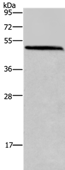
ApplicationsWestern Blot, ELISA
Product group Antibodies
ReactivityHuman
Overview
- SupplierApplied Biological Materials
- Product NameAnti-HOMER1
- Delivery Days Customer9
- ApplicationsWestern Blot, ELISA
- CertificationResearch Use Only
- ClonalityPolyclonal
- HostRabbit
- Scientific DescriptionThis gene encodes a member of the homer family of dendritic proteins. Members of this family regulate group 1 metabotrophic glutamate receptor function. [provided by RefSeq, Jul 2008]
- ReactivityHuman
- Storage Instruction-20°C,2°C to 8°C
- UNSPSC12352203
Related products
Product group Antibodies
Anti-HOMER1 AntibodyA37225
ApplicationsWestern Blot, ImmunoHistoChemistry
ReactivityHuman, Mouse
- SizePrice
Product group Antibodies
Anti-HOMER1 Antibody101-10728
ApplicationsWestern Blot, ELISA
TargetHOMER1
- SizePrice

Product group Antibodies
Anti-HOMER1 Antibody Picoband(r)A03877-1-CARRIER-FREE
ApplicationsFlow Cytometry, Western Blot, ELISA
ReactivityHuman, Mouse, Rat
TargetHOMER1
- SizePrice
Product group Antibodies
ApplicationsFlow Cytometry, Western Blot, ImmunoCytoChemistry
ReactivityHuman, Mouse, Rat
TargetHOMER1
- SizePrice
Product group Antibodies
HOMER1 AntibodyCSB-PA03007A0RB
ApplicationsELISA, ImmunoHistoChemistry
ReactivityHuman
TargetHOMER1
- SizePrice
Product group Antibodies

HOMER1 / Homer 1 AntibodyLS-C401742
ApplicationsWestern Blot, ELISA, ImmunoHistoChemistry
ReactivityHuman, Mouse, Rat
TargetHOMER1
- SizePrice
![Homer1 antibody detects Homer1 protein by immunofluorescent analysis. Sample: DIV9 rat E18 primary cortical neurons were fixed in 4% paraformaldehyde at RT for 15 min. Green: Homer1 protein stained by Homer1 antibody (GTX103278) diluted at 1:500. Red: beta Tubulin 3/ Tuj1, stained by beta Tubulin 3/ Tuj1 antibody [GT886] (GTX631830) diluted at 1:500. Blue: Fluoroshield with DAPI (GTX30920).](https://www.genetex.com/upload/website/prouct_img/normal/GTX103278/GTX103278_39967_20170705_IFA_R_w_23060119_440.webp)
Product group Antibodies
Homer1 antibodyGTX103278
ApplicationsImmunoFluorescence, ImmunoPrecipitation, Western Blot, ImmunoCytoChemistry, ImmunoHistoChemistry, ImmunoHistoChemistry Frozen, ImmunoHistoChemistry Paraffin
ReactivityHuman, Mouse, Rat
TargetHOMER1
- SizePrice

Product group Antibodies
Anti-HOMER1 AntibodyHPA036522
ApplicationsWestern Blot, ImmunoCytoChemistry, ImmunoHistoChemistry
ReactivityHuman
TargetHOMER1
- SizePrice