


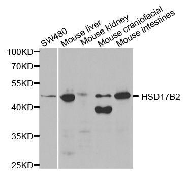
Figure 1. Western blot analysis of HSD17B2 using anti-HSD17B2 antibody (A02506-1). Electrophoresis was performed on a 5-20% SDS-PAGE gel at 70V (Stacking gel) / 90V (Resolving gel) for 2-3 hours. The sample well of each lane was loaded with 30 ug of sample under reducing conditions. Lane 1: human MCF-7 whole cell lysates, Lane 2: human RT4 whole cell lysates, Lane 3: human HepG2 whole cell lysates, Lane 4: human placenta tissue lysates, Lane 5: mouse liver tissue tissue lysates. After electrophoresis, proteins were transferred to a nitrocellulose membrane at 150 mA for 50-90 minutes. Blocked the membrane with 5% non-fat milk/TBS for 1.5 hour at RT. The membrane was incubated with rabbit anti-HSD17B2 antigen affinity purified polyclonal antibody (Catalog # A02506-1) at 0.5 microg/mL overnight at 4°C, then washed with TBS-0.1%Tween 3 times with 5 minutes each and probed with a goat anti-rabbit IgG-HRP secondary antibody at a dilution of 1:5000 for 1.5 hour at RT. The signal is developed using an Enhanced Chemiluminescent detection (ECL) kit (Catalog # EK1002) with Tanon 5200 system. A specific band was detected for HSD17B2 at approximately 37 kDa. The expected band size for HSD17B2 is at 43-49,35-40 kDa.
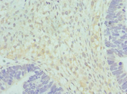
. HSD17B2 was detected in an immunocytochemical section of A431 cells. Enzyme antigen retrieval was performed using IHC enzyme antigen retrieval reagent (AR0022) for 15 mins. The cells were blocked with 10% goat serum. And then incubated with 5 microg/mL rabbit anti-HSD17B2 Antibody (A02506-1) overnight at 4°C. Cy3 Conjugated Goat Anti-Rabbit IgG (BA1032) was used as secondary antibody at 1:500 dilution and incubated for 30 minutes at 37°C. The section was counterstained with DAPI. Visualize using a fluorescence microscope and filter sets appropriate for the label used.")
. Overlay histogram showing MCF-7 cells stained with A02506-1 (Blue line). To facilitate intracellular staining, cells were fixed with 4% paraformaldehyde and permeabilized with permeabilization buffer. The cells were blocked with 10% normal goat serum. And then incubated with rabbit anti-HSD17B2 Antibody (A02506-1, 1 microg/1x106 cells) for 30 min at 20°C. DyLight®488 conjugated goat anti-rabbit IgG (BA1127, 5-10 microg/1x106 cells) was used as secondary antibody for 30 minutes at 20°C. Isotype control antibody (Green line) was rabbit IgG (1 microg/1x106) used under the same conditions. Unlabelled sample (Red line) was also used as a control.")
Figure 1. Western blot analysis of HSD17B2 using anti-HSD17B2 antibody (A02506-1). Electrophoresis was performed on a 5-20% SDS-PAGE gel at 70V (Stacking gel) / 90V (Resolving gel) for 2-3 hours. The sample well of each lane was loaded with 30 ug of sample under reducing conditions. Lane 1: human MCF-7 whole cell lysates, Lane 2: human RT4 whole cell lysates, Lane 3: human HepG2 whole cell lysates, Lane 4: human placenta tissue lysates, Lane 5: mouse liver tissue tissue lysates. After electrophoresis, proteins were transferred to a nitrocellulose membrane at 150 mA for 50-90 minutes. Blocked the membrane with 5% non-fat milk/TBS for 1.5 hour at RT. The membrane was incubated with rabbit anti-HSD17B2 antigen affinity purified polyclonal antibody (Catalog # A02506-1) at 0.5 microg/mL overnight at 4°C, then washed with TBS-0.1%Tween 3 times with 5 minutes each and probed with a goat anti-rabbit IgG-HRP secondary antibody at a dilution of 1:5000 for 1.5 hour at RT. The signal is developed using an Enhanced Chemiluminescent detection (ECL) kit (Catalog # EK1002) with Tanon 5200 system. A specific band was detected for HSD17B2 at approximately 37 kDa. The expected band size for HSD17B2 is at 43-49,35-40 kDa.
Anti-HSD17B2 Antibody Picoband(r)
A02506-1-CARRIER-FREE
ApplicationsFlow Cytometry, ImmunoFluorescence, Western Blot, ELISA, ImmunoCytoChemistry
Product group Antibodies
ReactivityHuman, Mouse
TargetHSD17B2
Overview
- SupplierBoster Bio
- Product NameAnti-HSD17B2 Antibody Picoband(r)
- Delivery Days Customer9
- Application Supplier NoteTested Species: In-house tested species with positive results. Other applications have not been tested. Optimal dilutions should be determined by end users.
- ApplicationsFlow Cytometry, ImmunoFluorescence, Western Blot, ELISA, ImmunoCytoChemistry
- CertificationResearch Use Only
- ClonalityPolyclonal
- Concentration500 ug/ml
- Gene ID3294
- Target nameHSD17B2
- Target descriptionhydroxysteroid 17-beta dehydrogenase 2
- Target synonymsEDH17B2, HSD17, SDR9C2, 17-beta-hydroxysteroid dehydrogenase type 2, 17-beta-HSD 2, 20 alpha-hydroxysteroid dehydrogenase, 20-alpha-HSD, E2DH, estradiol 17-beta-dehydrogenase 2, microsomal 17-beta-hydroxysteroid dehydrogenase, short chain dehydrogenase/reductase family 9C member 2, testosterone 17-beta-dehydrogenase
- HostRabbit
- IsotypeIgG
- Protein IDP37059
- Protein Name17-beta-hydroxysteroid dehydrogenase type 2
- Scientific DescriptionBoster Bio Anti-HSD17B2 Antibody Picoband® catalog # A02506-1. Tested in ELISA, IF, ICC, WB, Flow Cytometry applications. This antibody reacts with Human, Mouse. The brand Picoband indicates this is a premium antibody that guarantees superior quality, high affinity, and strong signals with minimal background in Western blot applications. Only our best-performing antibodies are designated as Picoband, ensuring unmatched performance.
- ReactivityHuman, Mouse
- Storage Instruction-20°C,2°C to 8°C
- UNSPSC12352203
Related products
Product group Antibodies
Anti-HSD17B2 Antibody144-01983
ApplicationsWestern Blot, ImmunoHistoChemistry
ReactivityHuman, Mouse, Rat
TargetHSD17B2
- SizePrice
Product group Antibodies
Anti-HSD17B2 AntibodyA34857
ApplicationsImmunoFluorescence, Western Blot, ImmunoHistoChemistry
ReactivityHuman
- SizePrice

Product group Antibodies
HSD17B2 Polyclonal AntibodyBS-3856R
ApplicationsImmunoFluorescence, Western Blot, ELISA, ImmunoCytoChemistry, ImmunoHistoChemistry, ImmunoHistoChemistry Frozen, ImmunoHistoChemistry Paraffin
ReactivityHuman, Mouse, Rat
TargetHSD17B2
- SizePrice
Product group Antibodies
HSD17B2 AntibodyCSB-PA010772ESR1HU
ApplicationsELISA, ImmunoHistoChemistry
ReactivityHuman
TargetHSD17B2
- SizePrice
Product group Antibodies

HSD17B2 AntibodyLS-C401751
ApplicationsWestern Blot, ELISA, ImmunoHistoChemistry
ReactivityHuman
TargetHSD17B2
- SizePrice

Product group Antibodies
Anti-HSD17B2 AntibodyHPA021826
ApplicationsWestern Blot, ImmunoCytoChemistry, ImmunoHistoChemistry
ReactivityHuman
TargetHSD17B2
- SizePrice
Product group Antibodies
HSD17B2 antibodyGTX54097
ApplicationsWestern Blot
ReactivityHuman, Mouse, Rat
TargetHSD17B2
- SizePrice
Product group Antibodies
ApplicationsWestern Blot, ELISA
ReactivityHuman
TargetHSD17B2
- SizePrice