


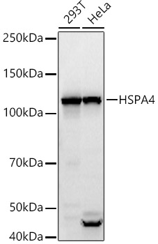
Figure 1. Western blot analysis of HSPA4 using anti-HSPA4 antibody (A03618-2). Electrophoresis was performed on a 5-20% SDS-PAGE gel at 70V (Stacking gel) / 90V (Resolving gel) for 2-3 hours. The sample well of each lane was loaded with 30 ug of sample under reducing conditions. Lane 1: human 293T whole cell lysates, Lane 2: human K562 whole cell lysates, Lane 3: rat brain tissue lysates, Lane 4: rat C6 whole cell lysates, Lane 6: mouse brain tissue lysates, Lane 7: mouse NIH/3T3 whole cell lysates, Lane 8: mouse Raw264.7 whole cell lysates. After electrophoresis, proteins were transferred to a nitrocellulose membrane at 150 mA for 50-90 minutes. Blocked the membrane with 5% non-fat milk/TBS for 1.5 hour at RT. The membrane was incubated with rabbit anti-HSPA4 antigen affinity purified polyclonal antibody (Catalog # A03618-2) at 0.25 microg/mL overnight at 4°C, then washed with TBS-0.1%Tween 3 times with 5 minutes each and probed with a goat anti-rabbit IgG-DyLight 647 Conjugated secondary antibody at a dilution of 1:2000 for 1.5 hour at RT. A specific band was detected for HSPA4 at approximately 110 kDa. The expected band size for HSPA4 is at 96,14 kDa.
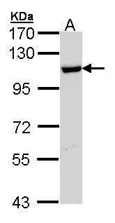
. Electrophoresis was performed on a 5-20% SDS-PAGE gel at 70V (Stacking gel) / 90V (Resolving gel) for 2-3 hours. The sample well of each lane was loaded with 30 ug of sample under reducing conditions. Lane 1: human 293T whole cell lysates, Lane 2: human K562 whole cell lysates, Lane 3: rat brain tissue lysates, Lane 4: rat C6 whole cell lysates, Lane 5: mouse brain tissue lysates, Lane 6: mouse NIH/3T3 whole cell lysates, Lane 7: mouse Raw264.7 whole cell lysates. After electrophoresis, proteins were transferred to a nitrocellulose membrane at 150 mA for 50-90 minutes. Blocked the membrane with 5% non-fat milk/TBS for 1.5 hour at RT. The membrane was incubated with rabbit anti-HSPA4 antigen affinity purified polyclonal antibody (Catalog # A03618-2) at 0.5 microg/mL overnight at 4°C, then washed with TBS-0.1%Tween 3 times with 5 minutes each and probed with a goat anti-rabbit IgG-HRP secondary antibody at a dilution of 1:5000 for 1.5 hour at RT. The signal is developed using an Enhanced Chemiluminescent detection (ECL) kit (Catalog # EK1002) with Tanon 5200 system. A specific band was detected for HSPA4 at approximately 110 kDa. The expected band size for HSPA4 is at 94,16 kDa.")
. HSPA4 was detected in a paraffin-embedded section of human colon cancer tissue. Heat mediated antigen retrieval was performed in EDTA buffer (pH 8.0, epitope retrieval solution). The tissue section was blocked with 10% goat serum. The tissue section was then incubated with 2 microg/ml rabbit anti-HSPA4 Antibody (A03618-2) overnight at 4°C. Peroxidase Conjugated Goat Anti-rabbit IgG was used as secondary antibody and incubated for 30 minutes at 37°C. The tissue section was developed using HRP Conjugated Rabbit IgG Super Vision Assay Kit (Catalog # SV0002) with DAB as the chromogen.")
. HSPA4 was detected in a paraffin-embedded section of mouse ovary tissue. Heat mediated antigen retrieval was performed in EDTA buffer (pH 8.0, epitope retrieval solution). The tissue section was blocked with 10% goat serum. The tissue section was then incubated with 2 microg/ml rabbit anti-HSPA4 Antibody (A03618-2) overnight at 4°C. Peroxidase Conjugated Goat Anti-rabbit IgG was used as secondary antibody and incubated for 30 minutes at 37°C. The tissue section was developed using HRP Conjugated Rabbit IgG Super Vision Assay Kit (Catalog # SV0002) with DAB as the chromogen.")
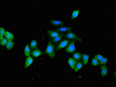
. Overlay histogram showing 293T cells stained with A03618-2 (Blue line). To facilitate intracellular staining, cells were fixed with 4% paraformaldehyde and permeabilized with permeabilization buffer. The cells were blocked with 10% normal goat serum. And then incubated with rabbit anti-HSPA4 Antibody (A03618-2, 1 microg/1x106 cells) for 30 min at 20°C. DyLight®488 conjugated goat anti-rabbit IgG (BA1127, 5-10 microg/1x106 cells) was used as secondary antibody for 30 minutes at 20°C. Isotype control antibody (Green line) was rabbit IgG (1 microg/1x106) used under the same conditions. Unlabelled sample without incubation with primary antibody and secondary antibody (Red line) was used as a blank control.")
. Overlay histogram showing MOLT-4 cells stained with A03618-2 (Blue line). To facilitate intracellular staining, cells were fixed with 4% paraformaldehyde and permeabilized with permeabilization buffer. The cells were blocked with 10% normal goat serum. And then incubated with rabbit anti-HSPA4 Antibody (A03618-2, 1 microg/1x106 cells) for 30 min at 20°C. DyLight®488 conjugated goat anti-rabbit IgG (BA1127, 5-10 microg/1x106 cells) was used as secondary antibody for 30 minutes at 20°C. Isotype control antibody (Green line) was rabbit IgG (1 microg/1x106) used under the same conditions. Unlabelled sample without incubation with primary antibody and secondary antibody (Red line) was used as a blank control.")
Figure 1. Western blot analysis of HSPA4 using anti-HSPA4 antibody (A03618-2). Electrophoresis was performed on a 5-20% SDS-PAGE gel at 70V (Stacking gel) / 90V (Resolving gel) for 2-3 hours. The sample well of each lane was loaded with 30 ug of sample under reducing conditions. Lane 1: human 293T whole cell lysates, Lane 2: human K562 whole cell lysates, Lane 3: rat brain tissue lysates, Lane 4: rat C6 whole cell lysates, Lane 6: mouse brain tissue lysates, Lane 7: mouse NIH/3T3 whole cell lysates, Lane 8: mouse Raw264.7 whole cell lysates. After electrophoresis, proteins were transferred to a nitrocellulose membrane at 150 mA for 50-90 minutes. Blocked the membrane with 5% non-fat milk/TBS for 1.5 hour at RT. The membrane was incubated with rabbit anti-HSPA4 antigen affinity purified polyclonal antibody (Catalog # A03618-2) at 0.25 microg/mL overnight at 4°C, then washed with TBS-0.1%Tween 3 times with 5 minutes each and probed with a goat anti-rabbit IgG-DyLight 647 Conjugated secondary antibody at a dilution of 1:2000 for 1.5 hour at RT. A specific band was detected for HSPA4 at approximately 110 kDa. The expected band size for HSPA4 is at 96,14 kDa.
Anti-HSPA4 Antibody Picoband(r)
A03618-2-CARRIER-FREE
ApplicationsFlow Cytometry, ImmunoFluorescence, Western Blot, ImmunoCytoChemistry, ImmunoHistoChemistry
Product group Antibodies
ReactivityHuman, Mouse, Rat
TargetHSPA4
Overview
- SupplierBoster Bio
- Product NameAnti-HSPA4 Antibody Picoband(r)
- Delivery Days Customer9
- ApplicationsFlow Cytometry, ImmunoFluorescence, Western Blot, ImmunoCytoChemistry, ImmunoHistoChemistry
- CertificationResearch Use Only
- ClonalityPolyclonal
- Concentration500 ug/ml
- Gene ID3308
- Target nameHSPA4
- Target descriptionheat shock protein family A (Hsp70) member 4
- Target synonymsAPG-2, HEL-S-5a, HS24/P52, HSPH2, RY, hsp70, hsp70RY, heat shock 70 kDa protein 4, epididymis secretory sperm binding protein Li 5a, heat shock 70-related protein APG-2, heat shock 70kD protein 4, heat shock protein family H member 2, heat shock protein, 110 kDa, hsp70 RY
- HostRabbit
- Protein IDP34932
- Protein NameHeat shock 70 kDa protein 4
- Scientific DescriptionBoster Bio Anti-HSPA4 Antibody Picoband® catalog # A03618-2. Tested in WB, IHC, ICC/IF, Flow Cytometry applications. This antibody reacts with Human, Mouse, Rat. The brand Picoband indicates this is a premium antibody that guarantees superior quality, high affinity, and strong signals with minimal background in Western blot applications. Only our best-performing antibodies are designated as Picoband, ensuring unmatched performance.
- ReactivityHuman, Mouse, Rat
- Storage Instruction-20°C,2°C to 8°C
- UNSPSC12352203
Related products
Product group Antibodies
Anti-HSPA4 AntibodyA308064
ApplicationsWestern Blot
ReactivityHuman, Mouse, Rat
- SizePrice
Product group Antibodies
Anti-HSPA4 Antibody101-10756
ApplicationsWestern Blot, ELISA
TargetHSPA4
- SizePrice
Product group Antibodies
HSPA4 Recombinant Antibody, AbBy Fluor-350 ConjugatedBSM-61343R-BF350
ApplicationsFlow Cytometry, ImmunoFluorescence, Western Blot
ReactivityHuman, Mouse, Rat
TargetHSPA4
- SizePrice
Product group Antibodies
HSPA4 AntibodyCSB-PA010825LA01HU
ApplicationsImmunoFluorescence, ELISA
ReactivityHuman
TargetHSPA4
- SizePrice
Product group Antibodies
ApplicationsImmunoPrecipitation, Western Blot, ImmunoCytoChemistry, ImmunoHistoChemistry
ReactivityMouse
TargetHSPA4
- SizePrice
Product group Antibodies

HSPA4 / APG-2 Antibody (HRP)LS-C499070
ApplicationsELISA
ReactivityHuman
TargetHSPA4
- SizePrice

Product group Antibodies
Anti-HSPA4 AntibodyHPA010023
ApplicationsWestern Blot, ImmunoCytoChemistry, ImmunoHistoChemistry
ReactivityHuman, Mouse, Rat
TargetHSPA4
- SizePrice
Product group Antibodies
HSPA4 antibody [N1N3]GTX112329
ApplicationsWestern Blot, ImmunoHistoChemistry, ImmunoHistoChemistry Paraffin
ReactivityHuman, Rat
TargetHSPA4
- SizePrice