


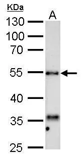
Figure 1. Western blot analysis of HYAL1 using anti-HYAL1 antibody (PA2181-1). Electrophoresis was performed on a 5-20% SDS-PAGE gel at 70V (Stacking gel) / 90V (Resolving gel) for 2-3 hours. The sample well of each lane was loaded with 30 ug of sample under reducing conditions. Lane 1: human HepG2 whole cell lysates, Lane 2: human PC-3 whole cell lysates, Lane 3: monkey COS-7 whole cell lysates, Lane 4: rat liver tissue lysates, Lane 5: mouse liver tissue lysates, Lane 6: mouse kidney tissue lysates. After electrophoresis, proteins were transferred to a nitrocellulose membrane at 150 mA for 50-90 minutes. Blocked the membrane with 5% non-fat milk/TBS for 1.5 hour at RT. The membrane was incubated with rabbit anti-HYAL1 antigen affinity purified polyclonal antibody (Catalog # PA2181-1) at 0.5 microg/mL overnight at 4°C, then washed with TBS-0.1%Tween 3 times with 5 minutes each and probed with a goat anti-rabbit IgG-HRP secondary antibody at a dilution of 1:5000 for 1.5 hour at RT. The signal is developed using an Enhanced Chemiluminescent detection (ECL) kit (Catalog # EK1002) with Tanon 5200 system. A specific band was detected for HYAL1 at approximately 60 kDa. The expected band size for HYAL1 is at 48 kDa.
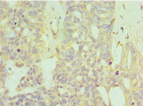
. HYAL1 was detected in a paraffin-embedded section of Human Intestinal Cancer tissue. Heat mediated antigen retrieval was performed in EDTA buffer (pH 8.0, epitope retrieval solution). The tissue section was blocked with 10% goat serum. The tissue section was then incubated with 1 microg/ml rabbit anti-HYAL1 Antibody (PA2181-1) overnight at 4°C. Peroxidase Conjugated Goat Anti-rabbit IgG was used as secondary antibody and incubated for 30 minutes at 37°C. The tissue section was developed using HRP Conjugated Rabbit IgG Super Vision Assay Kit (Catalog # SV0002) with DAB as the chromogen.")
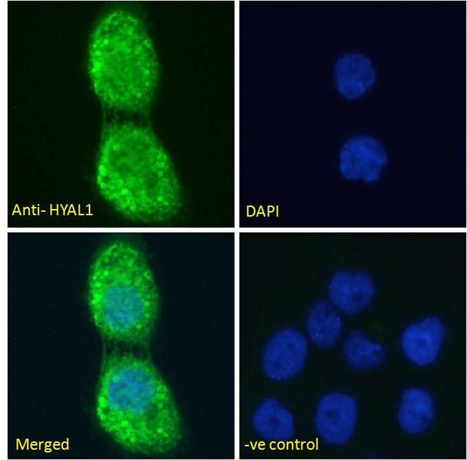
. HYAL1 was detected in an immunocytochemical section of MCF-7 cells. Enzyme antigen retrieval was performed using IHC enzyme antigen retrieval reagent (AR0022) for 15 mins. The cells were blocked with 10% goat serum. And then incubated with 1 microg/ml rabbit anti-HYAL1 Antibody (PA2181-1) overnight at 4°C. Biotinylated goat anti-rabbit IgG was used as secondary antibody and incubated for 30 minutes at 37°C. The section was developed using Strepavidin-Biotin-Complex (SABC)(Catalog # SA1022) with DAB as the chromogen.")
. HYAL1 was detected in a frozen section of Human Placenta tissue. The tissue section was blocked with 10% goat serum. The tissue section was then incubated with 1 microg/ml rabbit anti-HYAL1 Antibody (PA2181-1) overnight at 4°C. Biotinylated goat anti-rabbit IgG was used as secondary antibody and incubated for 30 minutes at 37°C. The tissue section was developed using Strepavidin-Biotin-Complex (SABC) (Catalog # SA1022) with DAB as the chromogen.")
Figure 1. Western blot analysis of HYAL1 using anti-HYAL1 antibody (PA2181-1). Electrophoresis was performed on a 5-20% SDS-PAGE gel at 70V (Stacking gel) / 90V (Resolving gel) for 2-3 hours. The sample well of each lane was loaded with 30 ug of sample under reducing conditions. Lane 1: human HepG2 whole cell lysates, Lane 2: human PC-3 whole cell lysates, Lane 3: monkey COS-7 whole cell lysates, Lane 4: rat liver tissue lysates, Lane 5: mouse liver tissue lysates, Lane 6: mouse kidney tissue lysates. After electrophoresis, proteins were transferred to a nitrocellulose membrane at 150 mA for 50-90 minutes. Blocked the membrane with 5% non-fat milk/TBS for 1.5 hour at RT. The membrane was incubated with rabbit anti-HYAL1 antigen affinity purified polyclonal antibody (Catalog # PA2181-1) at 0.5 microg/mL overnight at 4°C, then washed with TBS-0.1%Tween 3 times with 5 minutes each and probed with a goat anti-rabbit IgG-HRP secondary antibody at a dilution of 1:5000 for 1.5 hour at RT. The signal is developed using an Enhanced Chemiluminescent detection (ECL) kit (Catalog # EK1002) with Tanon 5200 system. A specific band was detected for HYAL1 at approximately 60 kDa. The expected band size for HYAL1 is at 48 kDa.
Anti-Hyaluronidase-1 HYAL1 Antibody Picoband(r)
PA2181-1-CARRIER-FREE
ApplicationsWestern Blot, ImmunoCytoChemistry, ImmunoHistoChemistry
Product group Antibodies
ReactivityHuman, Monkey, Mouse, Rat
TargetHYAL1
Overview
- SupplierBoster Bio
- Product NameAnti-Hyaluronidase-1 HYAL1 Antibody Picoband(r)
- Delivery Days Customer9
- ApplicationsWestern Blot, ImmunoCytoChemistry, ImmunoHistoChemistry
- CertificationResearch Use Only
- ClonalityPolyclonal
- Concentration500 ug/ml
- Gene ID3373
- Target nameHYAL1
- Target descriptionhyaluronidase 1
- Target synonymsHYAL-1, LUCA1, MPS9, NAT6, hyaluronidase-1, hyaluronoglucosaminidase 1, luCa-1, lung carcinoma protein 1, plasma hyaluronidase, tumor suppressor LUCA-1
- HostRabbit
- IsotypeIgG
- Protein IDQ12794
- Protein NameHyaluronidase-1
- Scientific DescriptionBoster Bio Anti-Hyaluronidase-1 HYAL1 Antibody catalog # PA2181-1. Tested in IHC, ICC, WB applications. This antibody reacts with Human, Monkey, Mouse, Rat. The brand Picoband indicates this is a premium antibody that guarantees superior quality, high affinity, and strong signals with minimal background in Western blot applications. Only our best-performing antibodies are designated as Picoband, ensuring unmatched performance.
- ReactivityHuman, Monkey, Mouse, Rat
- Storage Instruction-20°C,2°C to 8°C
- UNSPSC12352203
Related products
Product group Antibodies
HYAL1 AntibodyCSB-PA623651ESR1HU
ApplicationsELISA, ImmunoHistoChemistry
ReactivityHuman
TargetHYAL1
- SizePrice
Product group Antibodies
Anti-HYAL1 AntibodyA285977
ApplicationsFlow Cytometry, ImmunoFluorescence, ELISA, ImmunoHistoChemistry
ReactivityHuman
- SizePrice
Product group Antibodies

Goat anti-Hyaluronidase 1EB07871
ApplicationsFlow Cytometry, ImmunoFluorescence, ELISA, ImmunoHistoChemistry
ReactivityBovine, Canine, Human, Mouse, Porcine, Rat
TargetHYAL1
- SizePrice

Product group Antibodies
Anti-HYAL1 AntibodyHPA002112
ApplicationsImmunoHistoChemistry
ReactivityHuman
TargetHYAL1
- SizePrice
Product group Antibodies

HYAL1 AntibodyLS-C409001
ApplicationsWestern Blot, ImmunoHistoChemistry
ReactivityHuman, Mouse, Rat
TargetHYAL1
- SizePrice
Product group Antibodies
ApplicationsImmunoPrecipitation, Western Blot, ImmunoCytoChemistry, ImmunoHistoChemistry
TargetHYAL1
- SizePrice
Product group Antibodies
HYAL1 antibodyGTX113747
ApplicationsWestern Blot, ImmunoHistoChemistry, ImmunoHistoChemistry Paraffin
ReactivityHuman, Rat
TargetHYAL1
- SizePrice
Product group Antibodies
Anti-HYAL1Y058254
ApplicationsWestern Blot, ELISA, ImmunoHistoChemistry
ReactivityHuman
- SizePrice