


Immunohistochemical staining of human ovary shows cytoplasmic positivity in ovarian stroma cells.
![Lane 1: Marker [kDa] 250, 130, 95, 72, 55, 36, 28, 17, 10. Lane 2: Human cell line RT-4](https://atlasantibodies.s3.amazonaws.com/images/wb/hpa040092-wb-1.jpg "Lane 1: Marker [kDa] 250, 130, 95, 72, 55, 36, 28, 17, 10. Lane 2: Human cell line RT-4")
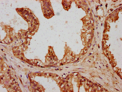
Immunohistochemical staining of human ovary shows cytoplasmic positivity in ovarian stroma cells.
Anti-HYI Antibody
HPA040092
ApplicationsWestern Blot, ImmunoCytoChemistry, ImmunoHistoChemistry
Product group Antibodies
ReactivityHuman
TargetHYI
Overview
- SupplierAtlas Antibodies
- Product NameAnti-HYI Antibody
- Delivery Days Customer4
- ApplicationsWestern Blot, ImmunoCytoChemistry, ImmunoHistoChemistry
- CertificationResearch Use Only
- ClonalityPolyclonal
- ConjugateUnconjugated
- Gene ID81888
- Target nameHYI
- Target descriptionhydroxypyruvate isomerase (putative)
- Target synonymsHT036, putative hydroxypyruvate isomerase, endothelial cell apoptosis protein E-CE1, hydroxypyruvate isomerase homolog
- HostRabbit
- IsotypeIgG
- Protein IDQ5T013
- Protein NamePutative hydroxypyruvate isomerase
- Scientific DescriptionRecombinant Protein Epitope Signature Tag (PrEST) antigen sequence
- ReactivityHuman
- Storage Instruction-20°C,2°C to 8°C
- UNSPSC41116161
Datasheet
MSDS
Related products

Product group Antibodies
Anti-HYI Antibody Picoband(r)A14191-2-CARRIER-FREE
ApplicationsFlow Cytometry, ImmunoFluorescence, Western Blot, ELISA, ImmunoCytoChemistry, ImmunoHistoChemistry
ReactivityHuman, Mouse, Rat
TargetHYI
- SizePrice
Product group Antibodies

HT036 / HYI AntibodyLS-C747878
ApplicationsWestern Blot
ReactivityHuman, Mouse, Rat
TargetHYI
- SizePrice
Product group Antibodies
HYI AntibodyCSB-PA719395LA01HU
ApplicationsELISA, ImmunoHistoChemistry
ReactivityHuman
TargetHYI
- SizePrice