


Immunofluorescent staining of human cell line U-2 OS shows localization to nucleoplasm.
Immunofluorescent staining of human cell line U-2 OS shows localization to nucleoplasm.
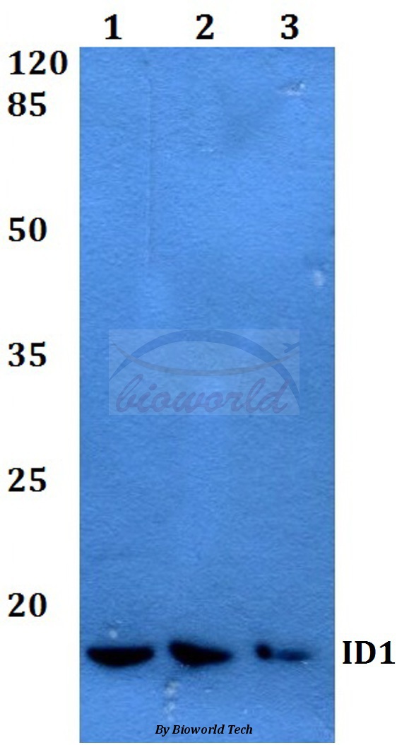
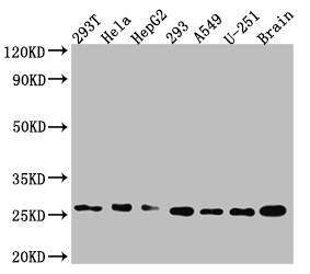
Anti-ID1 Antibody
HPA051456
ApplicationsImmunoCytoChemistry
Product group Antibodies
ReactivityHuman
TargetID1
Overview
- SupplierAtlas Antibodies
- Product NameAnti-ID1 Antibody
- Delivery Days Customer4
- ApplicationsImmunoCytoChemistry
- CertificationResearch Use Only
- ClonalityPolyclonal
- ConjugateUnconjugated
- Gene ID3397
- Target nameID1
- Target descriptioninhibitor of DNA binding 1
- Target synonymsID, bHLHb24, DNA-binding protein inhibitor ID-1, class B basic helix-loop-helix protein 24, dJ857M17.1.2 (inhibitor of DNA binding 1, dominant negative helix-loop-helix protein), inhibitor of DNA binding 1, HLH protein, inhibitor of DNA binding 1, dominant negative helix-loop-helix protein, inhibitor of differentiation 1
- HostRabbit
- IsotypeIgG
- Protein IDP41134
- Protein NameDNA-binding protein inhibitor ID-1
- Scientific DescriptionRecombinant Protein Epitope Signature Tag (PrEST) antigen sequence
- ReactivityHuman
- Storage Instruction-20°C,2°C to 8°C
- UNSPSC41116161
Datasheet
MSDS
Related products
Product group Antibodies
Anti-ID1 AntibodyA28242
ApplicationsWestern Blot
ReactivityHuman, Mouse, Rat
- SizePrice
Product group Antibodies
Anti-ID1 Antibody144-66610
ApplicationsWestern Blot
ReactivityHuman, Mouse, Rat
TargetID1
- SizePrice

Product group Antibodies
Anti-ID1 AntibodyAMAB91756
ApplicationsImmunoCytoChemistry, ImmunoHistoChemistry
ReactivityHuman
TargetID1
- SizePrice
Product group Antibodies
Anti-ID1 AntibodyAMAB91756
ApplicationsImmunoCytoChemistry, ImmunoHistoChemistry
ReactivityHuman
TargetID1
- SizePrice

Product group Antibodies
Anti-ID1 AntibodyAMAB91757
ApplicationsImmunoCytoChemistry, ImmunoHistoChemistry
ReactivityHuman
TargetID1
- SizePrice

Product group Antibodies
Id1 Recombinant AntibodyBSM-54101R
ApplicationsFlow Cytometry, ImmunoFluorescence, ImmunoCytoChemistry, ImmunoHistoChemistry, ImmunoHistoChemistry Frozen, ImmunoHistoChemistry Paraffin
ReactivityHuman
TargetID1
- SizePrice
Product group Antibodies
ID1 Recombinant Monoclonal AntibodyCSB-RA253477A0HU
ApplicationsImmunoFluorescence, Western Blot, ELISA
ReactivityHuman, Mouse
TargetID1
- SizePrice
Product group Antibodies
Id1 Recombinant AntibodyCAC10236
ApplicationsImmunoFluorescence, Western Blot, ELISA
ReactivityMouse
TargetID1
- SizePrice