


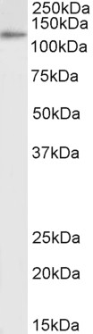
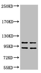
Figure 1. Western blot analysis of IDE using anti-IDE antibody (M01358). Electrophoresis was performed on a 5-20% SDS-PAGE gel at 70V (Stacking gel) / 90V (Resolving gel) for 2-3 hours. The sample well of each lane was loaded with 30 ug of sample under reducing conditions. Lane 1: human HepG2 whole cell lysates, Lane 2: human U251 whole cell lysates, Lane 3: human U2OS whole cell lysates, Lane 4: human SH-SY5Y whole cell lysates, Lane 5: rat brain tissue lysates, Lane 6: rat C6 whole cell lysates, Lane 7: mouse brain tissue lysates, Lane 8: mouse Neuro-2a whole cell lysates. After electrophoresis, proteins were transferred to a nitrocellulose membrane at 150 mA for 50-90 minutes. Blocked the membrane with 5% non-fat milk/TBS for 1.5 hour at RT. The membrane was incubated with rabbit anti-IDE antigen affinity purified monoclonal antibody (M01358) at 1:500 overnight at 4°C, then washed with TBS-0.1%Tween 3 times with 5 minutes each and probed with a goat anti-rabbit IgG-HRP secondary antibody at a dilution of 1:500 for 1.5 hour at RT. The signal is developed using an Enhanced Chemiluminescent detection (ECL) kit (Catalog # EK1002) with Tanon 5200 system. A specific band was detected for IDE at approximately 118 kDa. The expected band size for IDE is at 118 kDa.
Figure 1. Western blot analysis of IDE using anti-IDE antibody (M01358). Electrophoresis was performed on a 5-20% SDS-PAGE gel at 70V (Stacking gel) / 90V (Resolving gel) for 2-3 hours. The sample well of each lane was loaded with 30 ug of sample under reducing conditions. Lane 1: human HepG2 whole cell lysates, Lane 2: human U251 whole cell lysates, Lane 3: human U2OS whole cell lysates, Lane 4: human SH-SY5Y whole cell lysates, Lane 5: rat brain tissue lysates, Lane 6: rat C6 whole cell lysates, Lane 7: mouse brain tissue lysates, Lane 8: mouse Neuro-2a whole cell lysates. After electrophoresis, proteins were transferred to a nitrocellulose membrane at 150 mA for 50-90 minutes. Blocked the membrane with 5% non-fat milk/TBS for 1.5 hour at RT. The membrane was incubated with rabbit anti-IDE antigen affinity purified monoclonal antibody (M01358) at 1:500 overnight at 4°C, then washed with TBS-0.1%Tween 3 times with 5 minutes each and probed with a goat anti-rabbit IgG-HRP secondary antibody at a dilution of 1:500 for 1.5 hour at RT. The signal is developed using an Enhanced Chemiluminescent detection (ECL) kit (Catalog # EK1002) with Tanon 5200 system. A specific band was detected for IDE at approximately 118 kDa. The expected band size for IDE is at 118 kDa.
Anti-IDE/Insulysin Rabbit Monoclonal Antibody
M01358
ApplicationsWestern Blot, ImmunoHistoChemistry
Product group Antibodies
ReactivityHuman, Mouse, Rat
TargetIDE
Overview
- SupplierBoster Bio
- Product NameAnti-IDE/Insulysin Rabbit Monoclonal Antibody
- Delivery Days Customer9
- ApplicationsWestern Blot, ImmunoHistoChemistry
- CertificationResearch Use Only
- ClonalityMonoclonal
- Clone IDAOOB-9
- Gene ID3416
- Target nameIDE
- Target descriptioninsulin degrading enzyme
- Target synonymsINSULYSIN, insulin-degrading enzyme, Abeta-degrading protease, insulin protease, insulinase
- HostRabbit
- IsotypeIgG
- Protein IDP14735
- Protein NameInsulin-degrading enzyme
- Scientific DescriptionBoster Bio Anti-IDE/Insulysin Rabbit Monoclonal Antibody catalog # M01358. Tested in WB, IHC applications. This antibody reacts with Human, Mouse, Rat.
- ReactivityHuman, Mouse, Rat
- Storage Instruction-20°C
- UNSPSC12352203
Datasheet
MSDS
Related products
Product group Antibodies
ApplicationsWestern Blot, ELISA
ReactivityHuman
- SizePrice
Product group Antibodies

Anti-Insulin-degrading Enzyme [Fab-IDE]AB00406-1.1-BT
ApplicationsELISA
ReactivityHuman
TargetIDE
- SizePrice
Product group Antibodies
Anti-IDE Antibody144-01630
ApplicationsImmunoFluorescence, Western Blot, ImmunoHistoChemistry
ReactivityHuman, Mouse
TargetIDE
- SizePrice
Product group Antibodies

Insulysin AntibodyABX430884
ApplicationsWestern Blot, ELISA, ImmunoHistoChemistry
- SizePrice
Product group Antibodies

IDE Antibody (clone 1H4)LS-C767393
ApplicationsImmunoFluorescence, Western Blot, ImmunoHistoChemistry, ImmunoHistoChemistry Paraffin
ReactivityHuman
TargetIDE
- SizePrice
Product group Antibodies
IDE Recombinant Antibody, Biotin ConjugatedBSM-61379R-BIOTIN
ApplicationsWestern Blot, ImmunoHistoChemistry, ImmunoHistoChemistry Frozen, ImmunoHistoChemistry Paraffin
ReactivityHuman, Mouse, Rat
TargetIDE
- SizePrice
Product group Antibodies
IDE Monoclonal AntibodyCSB-MA000241
ApplicationsWestern Blot, ELISA
ReactivityHuman
TargetIDE
- SizePrice
Product group Antibodies

ApplicationsWestern Blot, ELISA, ImmunoHistoChemistry
ReactivityCanine, Human
TargetIDE
- SizePrice
Product group Antibodies
ApplicationsWestern Blot, ImmunoHistoChemistry
TargetIDE
- SizePrice