Bio-Connect
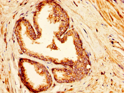
Immunohistochemical staining of human Endometrium shows moderate cytoplasmic positivity in glandular cells.

Anti-IDUA Antibody

HPA046979
Atlas Antibodies
ApplicationsWestern Blot, ImmunoHistoChemistry
Product group Antibodies
ReactivityHuman
TargetIDUA
Sign in to order and to see your custom pricing.
Large volume orders?
Order with a bulk request

Overview

  • Supplier
    Atlas Antibodies
  • Product Name
    Anti-IDUA
  • Delivery Days Customer
    4
  • Applications
    Western Blot, ImmunoHistoChemistry
  • Certification
    Research Use Only
  • Clonality
    Polyclonal
  • Conjugate
    Unconjugated
  • Gene ID3425
  • Target name
    IDUA
  • Target description
    alpha-L-iduronidase
  • Target synonyms
    alpha-L-iduronidase; IDA; iduronidase alpha-L-; MPS1; MPSI; mucopolysaccharidosis type I
  • Host
    Rabbit
  • Isotype
    IgG
  • Protein IDP35475
  • Protein Name
    Alpha-L-iduronidase
  • Scientific Description
    Recombinant Protein Epitope Signature Tag (PrEST) antigen sequence
  • Reactivity
    Human
  • Storage Instruction
    -20°C,2°C to 8°C
  • UNSPSC
    41116161

Related products

Product group Antibodies
RayBiotech
ApplicationsWestern Blot
TargetIDUA
  • SizePrice
Product group Antibodies
Biomatik
ApplicationsELISA, ImmunoHistoChemistry
TargetIDUA
  • SizePrice
IHC image of CSB-PA011000LA01HU diluted at 1:300 and staining in paraffin-embedded human prostate cancer performed on a Leica BondTM system. After dewaxing and hydration, antigen retrieval was mediated by high pressure in a citrate buffer (pH 6.0). Section was blocked with 10% normal goat serum 30min at RT. Then primary antibody (1% BSA) was incubated at 4°C overnight. The primary is detected by a biotinylated secondary antibody and visualized using an HRP conjugated SP system.
Product group Antibodies
Cusabio
IDUA AntibodyCSB-PA011000LA01HU
ApplicationsELISA, ImmunoHistoChemistry
ReactivityHuman
TargetIDUA
  • SizePrice
Immunohistochemistry analysis in human stomach and skeletal muscle tissues using HPA054254 antibody. Corresponding IDUA RNA-seq data are presented for the same tissues.
Product group Antibodies
Atlas Antibodies
ApplicationsImmunoHistoChemistry
ReactivityHuman
TargetIDUA
  • SizePrice
Immunohistochemistry analysis in human stomach and skeletal muscle tissues using HPA054254 antibody. Corresponding IDUA RNA-seq data are presented for the same tissues.
Product group Antibodies
Atlas Antibodies
ApplicationsImmunoHistoChemistry
ReactivityHuman
TargetIDUA
  • SizePrice
Figure 1. Western blot analysis of IDUA using anti-IDUA antibody (A02281-2). Electrophoresis was performed on a 5-20% SDS-PAGE gel at 70V (Stacking gel) / 90V (Resolving gel) for 2-3 hours. The sample well of each lane was loaded with 30 ug of sample under reducing conditions. Lane 1: human Hela whole cell lysates, Lane 2: human 293T whole cell lysates, Lane 3: human A549 whole cell lysates, Lane 4: human U87 whole cell lysates, Lane 5: rat brain tissue lysates, Lane 6: rat kidney tissue lysates, Lane 7: mouse brain tissue lysates, Lane 8: mouse kidney tissue lysates. After electrophoresis, proteins were transferred to a nitrocellulose membrane at 150 mA for 50-90 minutes. Blocked the membrane with 5% non-fat milk/TBS for 1.5 hour at RT. The membrane was incubated with rabbit anti-IDUA antigen affinity purified polyclonal antibody (Catalog # A02281-2) at 0.5 microg/mL overnight at 4°C, then washed with TBS-0.1%Tween 3 times with 5 minutes each and probed with a goat anti-rabbit IgG-HRP secondary antibody at a dilution of 1:5000 for 1.5 hour at RT. The signal is developed using an Enhanced Chemiluminescent detection (ECL) kit (Catalog # EK1002) with Tanon 5200 system. A specific band was detected for IDUA at approximately 73 kDa. The expected band size for IDUA is at 73 kDa.
Product group Antibodies
Boster Bio
Anti-IDUA Antibody Picoband(r)A02281-2-CARRIER-FREE
ApplicationsFlow Cytometry, ImmunoFluorescence, Western Blot, ELISA, ImmunoHistoChemistry
TargetIDUA
  • SizePrice
Formalin-fixed and paraffin embedded rat brain tissue probed with Anti-IDUA Polyclonal Antibody (bs-15542R) at a dilution of 1:200 overnight at 4°C, followed by Goat Anti-Rabbit secondary, Cy3 conjugated (bs-0295G-Cy3) at 1:200 dilution for 40 minutes at 37°C. DAPI(5ug/ml,blue,C-0033) was used to stain the cell nuclei
Product group Antibodies
Bioss Antibodies
ApplicationsWestern Blot, ELISA
TargetIDUA
  • SizePrice
Product group Antibodies
LifeSpan BioSciences
ApplicationsELISA, ImmunoHistoChemistry
TargetIDUA
  • SizePrice
Atlas Antibodies
Protein IDP35475
  • SizePrice
Atlas Antibodies
Protein IDP35475
  • SizePrice