Bio-Connect
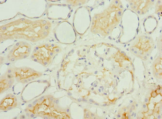
Immunohistochemical staining of human kidney shows moderate cytoplasmic positivity in cells in tubules.

Anti-IGFL3 Antibody

HPA062776
Atlas Antibodies
ApplicationsImmunoHistoChemistry
Product group Antibodies
TargetIGFL3
Sign in to order and to see your custom pricing.
Large volume orders?
Order with a bulk request

Overview

  • Supplier
    Atlas Antibodies
  • Product Name
    Anti-IGFL3
  • Delivery Days Customer
    4
  • Applications
    ImmunoHistoChemistry
  • Certification
    Research Use Only
  • Clonality
    Polyclonal
  • Conjugate
    Unconjugated
  • Gene ID388555
  • Target name
    IGFL3
  • Target description
    IGF like family member 3
  • Target synonyms
    insulin growth factor-like family member 3; RPRC483; UNQ483
  • Host
    Rabbit
  • Isotype
    IgG
  • Protein IDQ6UXB1
  • Protein Name
    Insulin growth factor-like family member 3
  • Scientific Description
    Recombinant Protein Epitope Signature Tag (PrEST) antigen sequence
  • Storage Instruction
    -20°C,2°C to 8°C
  • UNSPSC
    12352203

Related products

Immunohistochemistry of paraffin-embedded human kidney tissue using CSB-PA757792ESR1HU at dilution of 1:100
Product group Antibodies
Cusabio
IGFL3 AntibodyCSB-PA757792ESR1HU
ApplicationsELISA, ImmunoHistoChemistry
TargetIGFL3
  • SizePrice
Product group Antibodies
Assay Genie
TargetIGFL3
  • SizePrice
Product group Antibodies
Biomatik
ApplicationsELISA, ImmunoHistoChemistry
TargetIGFL3
  • SizePrice
Anti-IGFL3 Antibody
Product group Antibodies
Antibodies.com
ApplicationsWestern Blot
  • SizePrice
Atlas Antibodies
Protein IDQ6UXB1
  • SizePrice
Figure 1. Western blot analysis of IGFL3 using anti-IGFL3 antibody (A16075-1). Electrophoresis was performed on a 5-20% SDS-PAGE gel at 70V (Stacking gel) / 90V (Resolving gel) for 2-3 hours. The sample well of each lane was loaded with 30 ug of sample under reducing conditions. Lane 1: human placenta tissue lysates. After electrophoresis, proteins were transferred to a nitrocellulose membrane at 150 mA for 50-90 minutes. Blocked the membrane with 5% non-fat milk/TBS for 1.5 hour at RT. The membrane was incubated with rabbit anti-IGFL3 antigen affinity purified polyclonal antibody (Catalog # A16075-1) at 0.5 microg/mL overnight at 4°C, then washed with TBS-0.1%Tween 3 times with 5 minutes each and probed with a goat anti-rabbit IgG-HRP secondary antibody at a dilution of 1:5000 for 1.5 hour at RT. The signal is developed using an Enhanced Chemiluminescent detection (ECL) kit (Catalog # EK1002) with Tanon 5200 system. A specific band was detected for IGFL3 at approximately 18 kDa. The expected band size for IGFL3 is at 14 kDa.
Product group Antibodies
Boster Bio
Anti-IGFL3 Antibody Picoband(r)A16075-1-CARRIER-FREE
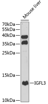
ApplicationsWestern Blot, ELISA, ImmunoHistoChemistry
TargetIGFL3
  • SizePrice
Product group Antibodies
LifeSpan BioSciences
IGFL3 AntibodyLS-C409129
ApplicationsWestern Blot, ImmunoHistoChemistry
TargetIGFL3
  • SizePrice