


Immunohistochemical staining of human rectum shows strong cytoplasmic positivity in glandular cells.
 and IQCE over-expression lysate (Co-expressed with a C-terminal myc-DDK tag (~3.1 kDa) in mammalian HEK293T cells, LY407468).")
Immunohistochemical staining of human rectum shows strong cytoplasmic positivity in glandular cells.
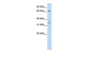
Anti-IQCE Antibody
HPA019515
ApplicationsWestern Blot, ImmunoCytoChemistry, ImmunoHistoChemistry
Product group Antibodies
ReactivityHuman
TargetIQCE
Overview
- SupplierAtlas Antibodies
- Product NameAnti-IQCE Antibody
- Delivery Days Customer4
- ApplicationsWestern Blot, ImmunoCytoChemistry, ImmunoHistoChemistry
- CertificationResearch Use Only
- ClonalityPolyclonal
- ConjugateUnconjugated
- Gene ID23288
- Target nameIQCE
- Target descriptionIQ motif containing E
- Target synonyms1700028P05Rik, PAPA7, IQ domain-containing protein E
- HostRabbit
- IsotypeIgG
- Protein IDQ6IPM2
- Protein NameIQ domain-containing protein E
- Scientific DescriptionRecombinant Protein Epitope Signature Tag (PrEST) antigen sequence
- ReactivityHuman
- Storage Instruction-20°C,2°C to 8°C
- UNSPSC41116161
Datasheet
MSDS
Related products

Product group Antibodies
Anti-IQCE Antibody Picoband(r)A13989-1-CARRIER-FREE
ApplicationsImmunoFluorescence, Western Blot, ELISA, ImmunoCytoChemistry
ReactivityHuman, Mouse, Rat
TargetIQCE
- SizePrice
Product group Antibodies

IQCE Antibody (aa385-545)LS-C178643
ApplicationsWestern Blot, ELISA, ImmunoHistoChemistry
ReactivityHuman
TargetIQCE
- SizePrice
Product group Antibodies
IQCE antibody, InternalGTX45268
ApplicationsWestern Blot
ReactivityHuman
TargetIQCE
- SizePrice
Product group Antibodies
Anti-IQCEY058881
ApplicationsWestern Blot, ELISA, ImmunoHistoChemistry
ReactivityHuman
- SizePrice
Product group Antibodies
IQCE Polyclonal AntibodyBS-9018R
ApplicationsImmunoFluorescence, Western Blot, ImmunoHistoChemistry, ImmunoHistoChemistry Paraffin
ReactivityHuman, Mouse, Rat
TargetIQCE
- SizePrice