

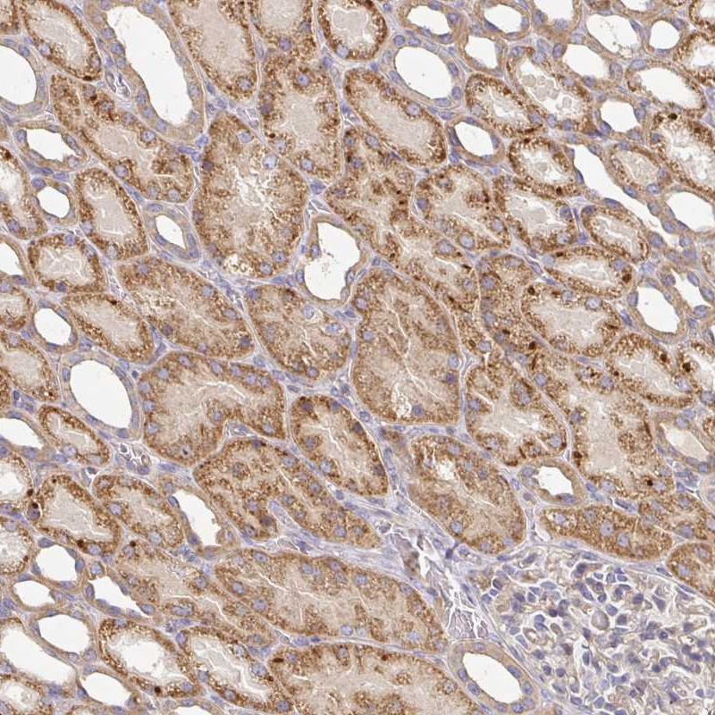
Immunohistochemical staining of human kidney shows moderate cytoplasmic positivity in cells in tubules.

Immunohistochemical staining of human kidney shows moderate cytoplasmic positivity in cells in tubules.
Anti-IRAK2 Antibody
HPA050520
ApplicationsImmunoCytoChemistry, ImmunoHistoChemistry
Product group Antibodies
ReactivityHuman
TargetIRAK2
Overview
- SupplierAtlas Antibodies
- Product NameAnti-IRAK2 Antibody
- Delivery Days Customer4
- ApplicationsImmunoCytoChemistry, ImmunoHistoChemistry
- CertificationResearch Use Only
- ClonalityPolyclonal
- ConjugateUnconjugated
- Gene ID3656
- Target nameIRAK2
- Target descriptioninterleukin 1 receptor associated kinase 2
- Target synonymsIRAK-2, interleukin-1 receptor-associated kinase-like 2
- HostRabbit
- IsotypeIgG
- Protein IDO43187
- Protein NameInterleukin-1 receptor-associated kinase-like 2
- Scientific DescriptionRecombinant Protein Epitope Signature Tag (PrEST) antigen sequence
- ReactivityHuman
- Storage Instruction-20°C,2°C to 8°C
- UNSPSC41116161
Datasheet
MSDS
Related products
Product group Antibodies
Anti-IRAK2 Antibody144-06635
ApplicationsImmunoFluorescence, Western Blot
ReactivityHuman, Mouse, Rat
TargetIRAK2
- SizePrice
Product group Antibodies

IRAK2 / IRAK-2 AntibodyLS-C831737
ApplicationsELISA, ImmunoHistoChemistry
ReactivityHuman
TargetIRAK2
- SizePrice

Product group Antibodies
Anti-IRAK2 Antibody Picoband(r)A01559-CARRIER-FREE
ApplicationsFlow Cytometry, ImmunoFluorescence, Western Blot, ImmunoCytoChemistry, ImmunoHistoChemistry
ReactivityHuman, Mouse, Rat
TargetIRAK2
- SizePrice
Product group Antibodies
IRAK2 AntibodyCSB-PA003062
ApplicationsWestern Blot, ELISA
ReactivityHuman
TargetIRAK2
- SizePrice
Product group Antibodies
ApplicationsImmunoPrecipitation, Western Blot, ImmunoCytoChemistry, ImmunoHistoChemistry
ReactivityMouse
TargetIRAK2
- SizePrice
Product group Antibodies
IRAK2 Polyclonal AntibodyBS-1427R
ApplicationsImmunoFluorescence, ELISA, ImmunoCytoChemistry, ImmunoHistoChemistry, ImmunoHistoChemistry Frozen, ImmunoHistoChemistry Paraffin
ReactivityHuman, Mouse, Rat
TargetIRAK2
- SizePrice
Product group Antibodies
IRAK2 antibody [N2C1], InternalGTX102518
ApplicationsImmunoFluorescence, Western Blot, ImmunoCytoChemistry
ReactivityHuman
TargetIRAK2
- SizePrice