

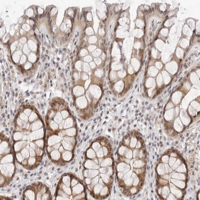
Immunohistochemical staining of human colon shows moderate nuclear and cytoplasmic positivity in glandular cells.
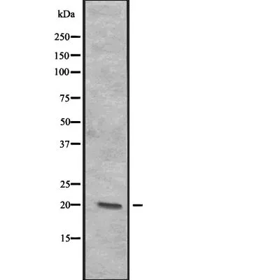
![Lane 1: Marker [kDa] 230, 130, 95, 72, 56, 36, 28, 17, 11. Lane 2: Human cell line RT-4. Lane 3: Human cell line U-251MG sp. Lane 4: Human plasma (IgG/HSA depleted). Lane 5: Human liver tissue. Lane 6: Human tonsil tissue](https://atlasantibodies.s3.amazonaws.com/images/wb/hpa028463-wb-1.jpg "Lane 1: Marker [kDa] 230, 130, 95, 72, 56, 36, 28, 17, 11. Lane 2: Human cell line RT-4. Lane 3: Human cell line U-251MG sp. Lane 4: Human plasma (IgG/HSA depleted). Lane 5: Human liver tissue. Lane 6: Human tonsil tissue")
Immunohistochemical staining of human colon shows moderate nuclear and cytoplasmic positivity in glandular cells.
Anti-ITGB3BP Antibody
HPA028463
ApplicationsWestern Blot, ImmunoCytoChemistry, ImmunoHistoChemistry
Product group Antibodies
ReactivityHuman
TargetITGB3BP
Overview
- SupplierAtlas Antibodies
- Product NameAnti-ITGB3BP Antibody
- Delivery Days Customer4
- ApplicationsWestern Blot, ImmunoCytoChemistry, ImmunoHistoChemistry
- CertificationResearch Use Only
- ClonalityPolyclonal
- ConjugateUnconjugated
- Gene ID23421
- Target nameITGB3BP
- Target descriptionintegrin subunit beta 3 binding protein
- Target synonymsCENP-R, CENPR, HSU37139, NRIF3, TAP20, centromere protein R, beta 3 endonexin, beta3-endonexin, integrin beta 3 binding protein (beta3-endonexin), integrin beta-3-binding protein, nuclear receptor-interacting factor 3
- HostRabbit
- IsotypeIgG
- Protein IDQ13352
- Protein NameCentromere protein R
- Scientific DescriptionRecombinant Protein Epitope Signature Tag (PrEST) antigen sequence
- ReactivityHuman
- Storage Instruction-20°C,2°C to 8°C
- UNSPSC41116161
Datasheet
MSDS
Related products
Product group Antibodies
ITGB3BP AntibodyCSB-PA622653DSR1HU
ApplicationsELISA, ImmunoHistoChemistry
ReactivityHuman
TargetITGB3BP
- SizePrice

Product group Antibodies
Anti-Centromere protein R/ITGB3BP Antibody Picoband(r)A09098-1-CARRIER-FREE
ApplicationsImmunoFluorescence, Western Blot, ELISA, ImmunoCytoChemistry
ReactivityHuman
TargetITGB3BP
- SizePrice
Product group Antibodies
Anti-ITGB3BP Antibody144-66009
ApplicationsWestern Blot
ReactivityHuman, Mouse, Rat
TargetITGB3BP
- SizePrice
Product group Antibodies
Anti-ITGB3BP AntibodyA91907
ApplicationsImmunoHistoChemistry
ReactivityHuman, Mouse, Rat
- SizePrice
Product group Antibodies

ITGB3BP AntibodyLS-C400825
ApplicationsELISA, ImmunoHistoChemistry
ReactivityHuman
TargetITGB3BP
- SizePrice
Product group Antibodies
ITGB3BP antibodyGTX03692
ApplicationsWestern Blot
ReactivityHuman
TargetITGB3BP
- SizePrice