


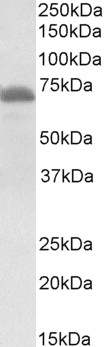
Figure 1. Western blot analysis of ITK using anti-ITK antibody (A01385-3). Electrophoresis was performed on a 5-20% SDS-PAGE gel at 70V (Stacking gel) / 90V (Resolving gel) for 2-3 hours. The sample well of each lane was loaded with 30 ug of sample under reducing conditions. Lane 1: human Jurkat whole cell lysates, Lane 2: human MOLT-4 whole cell lysates. After electrophoresis, proteins were transferred to a nitrocellulose membrane at 150 mA for 50-90 minutes. Blocked the membrane with 5% non-fat milk/TBS for 1.5 hour at RT. The membrane was incubated with rabbit anti-ITK antigen affinity purified polyclonal antibody (Catalog # A01385-3) at 0.5 microg/mL overnight at 4°C, then washed with TBS-0.1%Tween 3 times with 5 minutes each and probed with a goat anti-rabbit IgG-HRP secondary antibody at a dilution of 1:5000 for 1.5 hour at RT. The signal is developed using an Enhanced Chemiluminescent detection (ECL) kit (Catalog # EK1002) with Tanon 5200 system. A specific band was detected for ITK at approximately 72 kDa. The expected band size for ITK is at 72 kDa.
. ITK was detected in a paraffin-embedded section of human spleen cancer tissue. Heat mediated antigen retrieval was performed in EDTA buffer (pH 8.0, epitope retrieval solution). The tissue section was blocked with 10% goat serum. The tissue section was then incubated with 2 microg/ml rabbit anti-ITK Antibody (A01385-3) overnight at 4°C. Peroxidase Conjugated Goat Anti-rabbit IgG was used as secondary antibody and incubated for 30 minutes at 37°C. The tissue section was developed using HRP Conjugated Rabbit IgG Super Vision Assay Kit (Catalog # SV0002) with DAB as the chromogen.")
. Overlay histogram showing HEL cells stained with A01385-3 (Blue line). To facilitate intracellular staining, cells were fixed with 4% paraformaldehyde and permeabilized with permeabilization buffer. The cells were blocked with 10% normal goat serum. And then incubated with rabbit anti-ITK Antibody (A01385-3, 1 microg/1x106 cells) for 30 min at 20°C. DyLight®488 conjugated goat anti-rabbit IgG (BA1127, 5-10 microg/1x106 cells) was used as secondary antibody for 30 minutes at 20°C. Isotype control antibody (Green line) was rabbit IgG (1 microg/1x106) used under the same conditions. Unlabelled sample without incubation with primary antibody and secondary antibody (Red line) was used as a blank control.")
Figure 1. Western blot analysis of ITK using anti-ITK antibody (A01385-3). Electrophoresis was performed on a 5-20% SDS-PAGE gel at 70V (Stacking gel) / 90V (Resolving gel) for 2-3 hours. The sample well of each lane was loaded with 30 ug of sample under reducing conditions. Lane 1: human Jurkat whole cell lysates, Lane 2: human MOLT-4 whole cell lysates. After electrophoresis, proteins were transferred to a nitrocellulose membrane at 150 mA for 50-90 minutes. Blocked the membrane with 5% non-fat milk/TBS for 1.5 hour at RT. The membrane was incubated with rabbit anti-ITK antigen affinity purified polyclonal antibody (Catalog # A01385-3) at 0.5 microg/mL overnight at 4°C, then washed with TBS-0.1%Tween 3 times with 5 minutes each and probed with a goat anti-rabbit IgG-HRP secondary antibody at a dilution of 1:5000 for 1.5 hour at RT. The signal is developed using an Enhanced Chemiluminescent detection (ECL) kit (Catalog # EK1002) with Tanon 5200 system. A specific band was detected for ITK at approximately 72 kDa. The expected band size for ITK is at 72 kDa.
Anti-ITK Antibody Picoband(r)
A01385-3-CARRIER-FREE
ApplicationsFlow Cytometry, Western Blot, ELISA, ImmunoHistoChemistry
Product group Antibodies
ReactivityHuman
TargetITK
Overview
- SupplierBoster Bio
- Product NameAnti-ITK Antibody Picoband(r)
- Delivery Days Customer9
- ApplicationsFlow Cytometry, Western Blot, ELISA, ImmunoHistoChemistry
- CertificationResearch Use Only
- ClonalityPolyclonal
- Concentration500 ug/ml
- Gene ID3702
- Target nameITK
- Target descriptionIL2 inducible T cell kinase
- Target synonymsEMT, LPFS1, LYK, PSCTK2, tyrosine-protein kinase ITK/TSK, IL-2-inducible T-cell kinase, T-cell-specific kinase, homolog of mouse T-cell itk/tsk, interleukin-2-inducible T-cell kinase, kinase EMT, tyrosine-protein kinase LYK
- HostRabbit
- IsotypeIgG
- Protein IDQ08881
- Protein NameTyrosine-protein kinase ITK/TSK
- Scientific DescriptionBoster Bio Anti-ITK Antibody Picoband® catalog # A01385-3. Tested in ELISA, Flow Cytometry, IHC, WB applications. This antibody reacts with Human. The brand Picoband indicates this is a premium antibody that guarantees superior quality, high affinity, and strong signals with minimal background in Western blot applications. Only our best-performing antibodies are designated as Picoband, ensuring unmatched performance.
- ReactivityHuman
- Storage Instruction-20°C,2°C to 8°C
- UNSPSC12352203
Related products
Product group Antibodies
Anti-ITK/EMT AntibodyA84564
ApplicationsFlow Cytometry, ImmunoFluorescence, Western Blot, ELISA, ImmunoHistoChemistry
ReactivityHuman, Rat
- SizePrice
Product group Antibodies
Anti-Mouse Itk (N-term) Antibody102-23956
ApplicationsWestern Blot
TargetITK
- SizePrice
Product group Antibodies
ITK Recombinant Antibody, AbBy Fluor-594 ConjugatedBSM-61531R-BF594
ApplicationsFlow Cytometry, ImmunoFluorescence
ReactivityHuman
TargetITK
- SizePrice
Product group Antibodies
Phospho-ITK (Y512) AntibodyCSB-PA011277
ApplicationsWestern Blot, ELISA, ImmunoHistoChemistry
ReactivityHuman, Mouse
TargetITK
- SizePrice
Product group Antibodies

Goat anti-ITKEB05117
ApplicationsImmunoFluorescence, Western Blot, ELISA
ReactivityHuman, Mouse
TargetITK
- SizePrice
Product group Antibodies
Itk Recombinant AntibodyCAC12298
ApplicationsELISA, ImmunoHistoChemistry
TargetITK
- SizePrice
Product group Antibodies

ITK / EMT AntibodyLS-C405090
ApplicationsELISA, ImmunoHistoChemistry
ReactivityHuman, Mouse
TargetITK
- SizePrice

Product group Antibodies
Anti-ITK AntibodyHPA043670
ApplicationsImmunoCytoChemistry
ReactivityHuman
TargetITK
- SizePrice
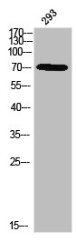
![Whole cell extract (30 μg) was separated by 7.5% SDS-PAGE, and the membrane was blotted with ITK antibody [N3C3] (GTX113215) diluted at 1:500. The HRP-conjugated anti-rabbit IgG antibody (GTX213110-01) was used to detect the primary antibody.](https://www.genetex.com/upload/website/prouct_img/normal/GTX113215/GTX113215_40128_20170810_WB_w_23060501_333.webp)
Product group Antibodies
ITK antibody [N3C3]GTX113215
ApplicationsWestern Blot
ReactivityHuman
TargetITK
- SizePrice